. 2017 Dec;7(12):170199.
doi: 10.1098/rsob.170199.

A eukaryotic nicotinate-inducible gene cluster: convergent evolution in fungi and bacteria

Affiliations

A eukaryotic nicotinate-inducible gene cluster: convergent evolution in fungi and bacteria

Judit Ámon et al. Open Biol. 2017 Dec.

Abstract

Nicotinate degradation has hitherto been elucidated only in bacteria. In the ascomycete Aspergillus nidulans, six loci, hxnS/AN9178 encoding the molybdenum cofactor-containing nicotinate hydroxylase, AN11197 encoding a Cys2/His2 zinc finger regulator HxnR, together with AN11196/hxnZ, AN11188/hxnY, AN11189/hxnP and AN9177/hxnT, are clustered and stringently co-induced by a nicotinate derivative and subject to nitrogen metabolite repression mediated by the GATA factor AreA. These genes are strictly co-regulated by HxnR. Within the hxnR gene, constitutive mutations map in two discrete regions. Aspergillus nidulans is capable of using nicotinate and its oxidation products 6-hydroxynicotinic acid and 2,5-dihydroxypyridine as sole nitrogen sources in an HxnR-dependent way. HxnS is highly similar to HxA, the canonical xanthine dehydrogenase (XDH), and has originated by gene duplication, preceding the origin of the Pezizomycotina. This cluster is conserved with some variations throughout the Aspergillaceae. Our results imply that a fungal pathway has arisen independently from bacterial ones. Significantly, the neo-functionalization of XDH into nicotinate hydroxylase has occurred independently from analogous events in bacteria. This work describes for the first time a gene cluster involved in nicotinate catabolism in a eukaryote and has relevance for the formation and evolution of co-regulated primary metabolic gene clusters and the microbial degradation of N-heterocyclic compounds.

Keywords: Cys2His2 transcription factor; convergent evolution; nicotinate catabolic gene cluster; nicotinate hydroxylase; xanthine dehydrogenase.


Conflict of interest statement

We have no competing interests.

Figures

Figure 1.
Figure 1.
Metabolic cross-talk between the purine and nicotinate utilization pathways. PHI is a conventional XDH able to catalyse the conversion of hypoxanthine to xanthine and xanthine to uric acid. XanA is an α-ketoglutarate-dependent xanthine dioxygenase, accepting xanthine but not hypoxanthine as a substrate. From there uric acid is converted into ammonium (formula image) by the well-established purine utilization pathway ([21] for review). PHII is an unconventional MOCO carrying enzyme hydroxylating hypoxanthine to xanthine and nicotinic acid to presumably 6-OH nicotinic acid. As this latter compound is a nitrogen source, it is presumably converted into ammonium, which is indicated by a dashed blue connector. Note that unlike PHI, PHII cannot use xanthine as a substrate. In black: steps induced by uric acid, under the control of the UaY transcription factor. In blue: steps actually (hxnS, PHII) or presumably induced by nicotinic acid, 6-OH nicotinic acid or a further metabolite in the nicotinate utilization pathway and under the control of the HxnR/AplA transcription factor(s). Full references are given in the text.
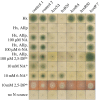
Figure 2.
Figure 2.
A comparison of PHI (HxA) and PHII (HxnS). An alignment of the two A. nidulans open reading frames with the structurally characterized XDH from B. taurus [24] is shown. Underlying the sequences: yellow, 2Fe/2S clusters; blue, FAD/NAD-binding domain; red, MOCO/substrate-binding subdomains I and II (as in [25]). Red arrows underlying the sequences indicate intron positions in the hxA gene, while green arrows indicate intron positions in hxnS. Boxed residues: yellow, conserved Cys in the 2Fe/2S clusters, also indicated the Glu45 and Gly46 (in B. taurus) residues belonging to the 2Fe/2S-binding loop, and separating this cluster from the flavin-binding ring; orange, FAD-binding residues [24]; blue, NAD+/NADH-interacting residues [26]; green, residues interacting with MOCO [25]; red, residues where HxnS and its putative orthologues differ from both HxA and typical XDHs represented by the B. taurus enzyme. Red asterisks mark residues involved in substrate binding of B. taurus XDH [24,27,28]. Blue asterisks mark residues lining the substrate access channel of B. taurus XDH [28]. Green asterisks mark residues hydrogen-bonding a molybdenum-bound oxygen [27]. Red downward arrows indicate mutational changes leading to complete loss of function in HxA; blue downward arrows indicate mutations leading to changes of substrate and inhibitor specificity in HxA [29]; the downward green arrow indicates the only extant missense mutation sequenced for HxnS. Alignment with MAFFT E-INS-i, visualized with BoxShade.
Figure 3.
Figure 3.
Utilization of different nitrogen sources by mutants described in this article. Above each column we indicate the relevant mutation carried by each tested strain. Hx indicates 1 mM hypoxanthine as the sole nitrogen source. Hx, Allp, as above including 5.5 µM allopurinol, which fully inhibits PHI (HxA) but not PHII (HxnS). NA, 6-NA and 2,5-DP indicate, respectively, nicotinic acid and 6-OH nicotinic acid added as the sodium salts (see ‘Material and methods’ section) and 2,5-dihydroxypyridine added as powder. Other relevant concentrations are indicated in the figure. Plates were incubated for 3 days at 37°C except those marked by asterisk (*), which were incubated for 4 days. Strains used: control 1 (HZS.120, parent of hxnSΔ), control 2 (TN02 A21) are wt for all hxn genes. Mutant strains: hxnSΔ (HZS.599), hxB20 (HZS.135), hxnRΔ (HZS.136), hxnR80 (HZS.220) and hxnRc7 (FGSC A872). The complete genotypes are given in the electronic supplementary material, table S5.
Figure 4.
Figure 4.
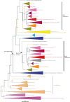
A simplified phylogeny of the fungal purine hydroxylases, HxA (PHI)-like and HxnS (PHII)-like. This tree in cartoon form was extracted from the more complete tree shown in the electronic supplementary material, figure S3, where all species used are indicated. Outgroups are the nearest non-fungal taxa of the Opisthokonta. Values at nodes are aLRTs (approximate likelihood ratio tests). The arrows indicate the putative nodes where the gene duplication and the PHII neo-functionalization occurred.
Figure 5.
Figure 5.
A schematic representation of the HxnR transcription factor and verification of constitutivity of hxnRc mutants. (a) A schematic of the HxnR transcription factor is shown, indicating the two Cys2His2 Zn-finger domains (C2H2, in purple), the putative nuclear localization signal (NLS, in orange), the putative nuclear export signal (NES, in yellow), the fungal transcription factor domain (pfam04082, in blue) and the two regions where the constitutive mutations occur (in green). The three extant loss-of-function mutations are indicated in red letters in the scheme. All the amino acid changes leading to constitutivity are indicated, together with the cognate allele number (in green). (b) Enzyme activity staining for hypoxanthine hydroxylase (Hx) and nicotinate hydroxylase (NA) of the constitutive mutants is shown. Only HxnS (PHII), which has a lower mobility than HxA (PHI), stains with nicotinate as the substrate. Note its complete absence in the wt strain hxnR+ hxA+ grown under non-inducing conditions, while HxA shows substantial basal levels as reported previously [1,38]. As the constitutive mutations were isolated in different hxA backgrounds (hxA18, hxAΔ, hxA+; see ‘Material and methods’ section and electronic supplementary material, table S2), this is also indicated. All mycelia were grown in non-inducing conditions (for either HxA or HxnS) on 1 mM acetamide as the nitrogen source for 15 h at 37°C.
Figure 6.
Figure 6.
Co-regulation of the genes in the hxn cluster. (a) mRNA levels measured by qRT-PCR for all the genes in the hxn cluster. Mycelia were grown on 1 mM acetamide as the sole nitrogen source for 8 h at 37°C. They were either maintained on the same medium for a further 2 h (non-induced, NI) or induced with 1 mM nicotinic acid (as the sodium salt, I) or induced as above together with 5 mM of l-(+)diammonium-tartrate (induced repressed, IR). Strains used: hxnR+ (FGSC A26), hxnRΔ (HZS.136) and hxnRc7 (FGSC A872). (b) Northern blot showing qualitatively the co-regulation of all the genes in the cluster under different growth conditions. Mycelia were grown on 500 µM urea for 8 h, and then transferred to 1 mM acetamide for an additional 2 h (non-induced, NI) or to the same plus 1 mM nicotinic acid (as above, I) or to the latter together with 5 mM l-(+)diammonium-tartrate (induced repressed, IR). Together with hxnS, hxnR, hxnT, hxnP, hxnY and hxnZ transcripts we also monitored the expression of hxB, an unlinked gene, which was previously shown to be under the control of HxnR [41]. As a loading control, the expression of acnA (actin) was monitored. Strains used are indicated by the relevant mutation: hxnR+ (FGSC A26), hxnR2 (CS302), a missense unleaky mutation (Gly76Asp) and hxnRc7 (FGSC A872), our standard constitutive mutation (figure 5; electronic supplementary material figure S4 and table S2). (c) Expression of hxnS and hxnP under conditions of nitrogen starvation. Mycelia were grown on 5 mM urea as the sole nitrogen source for 8 h, and then transferred to the same medium for two additional hours (U, which is non-inducing and actually partially repressed conditions; see text) or to a medium without any nitrogen source (starvation media, St) or to a medium with 10 mM nicotinic acid as the nitrogen source (inducing media, I). Strains as in panel (a). In all qRT-PCR experiments, data were processed according to the standard curve method with acnA as the control mRNA. Standard errors of three independent experiments are shown in all qRT-PCR. Gene probe primers are detailed in the electronic supplementary material, table S6. (d) Cluster arrangement of the hxn genes on chromosome VI.
Figure 7.
Figure 7.
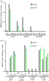
Induction depends on the AreA GATA factor and on the metabolism of nicotinic acid. (a) The GATA factor AreA is essential for hxnP and hxnS expression. hxnP and hxnS mRNA levels in areA+ (FGSC A26) and an areA supposedly derepressed mutant (xprD1, HZS.216) and areA null mutant (areA600, CS3095) strains. Non-induced conditions (NI): Strains were grown on MM media with 5 mM l-(+)diammonium-tartrate as the sole N-source for 8 h, and then the mycelia were transferred to MM with 1 mM acetamide for further 2 h. Induced conditions (I): as above but transferred to 10 mM nicotinic acid as the sole N-source. Induced repressed conditions (IR): transferred to 10 mM nicotinic acid and 5 mM diammonium-tartrate. N-starvation conditions (St): transferred to nitrogen-free medium. (b) Induction depends on metabolism of nicotinic acid via HxnS activity. mRNA levels of hxnP and hxnS in hxnR+ hxB+ (FGSC A26), hxnR+ hxB20 (HZS.135), hxnRΔ hxB+ (HZS.136) and hxnR constitutive, hxnRc7 hxB+ (FGSC A872) strains are shown. Non-induced (NI) and induced growth conditions were the same as detailed in (a). (NA): induced with 1 mM nicotinic acid; (6-NA): induced with 1 mM 6-OH nicotinic acid. The hxB20 mutation abolishes completely HxnS activity without affecting its expression as judged by measuring its CRM [12]. qRT-PCR data in both panels were processed according to the standard curve method; the housekeeping control transcript was actin (acnA). Standard deviations based on three biological replicates are shown.
Figure 8.
Figure 8.
Hxn cluster organization in the Aspergillaceae family. Boxes indicate genes; arrowheads indicate orientation. Colour stands for the orthologues found in different species (Aspergillus nidulans, A. niger, A. flavus, A. ruber, Monascus ruber, A. wentii, A. sclerotiorum, A. ochraceoroseus, A. fumigatus, Penicillium citrinum, P. digitatum). Stars indicate putative pseudogenes (putative non-functional alleles); hatched boxes indicate duplicated paralogues. Vertical lines symbolize physical unlinkage of genes on the same chromosomes. The blank box in A. niger stands for the orthologue of the A. nidulans gene at locus AN8360 (encoding a nitroreductase of bacterial origin), which is unlinked to the cluster in the latter fungus, while its expression is not regulated by nicotinate or the transcription factor HxnR.

References

    1. Scazzocchio C, Holl FB, Foguelman AI. 1973. The genetic control of molybdoflavoproteins in Aspergillus nidulans. Allopurinol-resistant mutants constitutive for xanthine-dehydrogenase. Eur. J. Biochem. 36, 428–445. (doi:10.1111/j.1432-1033.1973.tb02928.x) - DOI - PubMed
    1. Scazzocchio C. 1980. The genetics of the molybdenum containing enzymes. In Molybdenum and molybdenum-containing enzymes (ed. Coughlan MP.), pp. 487–515. Oxford, UK: Pergamon Press.
    1. Scazzocchio C. 1994. The purine degradation pathway, genetics, biochemistry and regulation. Prog. Ind. Microbiol. 29, 221–257. - PubMed
    1. Pateman JA, Cove DJ, Rever BM, Roberts DB. 1964. A common co-factor for nitrate reductase and xanthine dehydrogenase which also regulates the synthesis of nitrate reductase. Nature 201, 58–60. (doi:10.1038/201058a0) - DOI - PubMed
    1. Schwarz G, Mendel RR, Ribbe MW. 2009. Molybdenum cofactors, enzymes and pathways. Nature 460, 839–847. (doi:10.1038/nature08302) - DOI - PubMed

MeSH terms

LinkOut - more resources