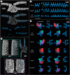
Water vascular system architecture in an Ordovician ophiuroid
- PMID: 29212753
- PMCID: PMC5746540
- DOI: 10.1098/rsbl.2017.0635
Water vascular system architecture in an Ordovician ophiuroid
Abstract
Understanding the water vascular system (WVS) in early fossil echinoderms is critical to elucidating the evolution of this system in extant forms. Here we present the first report of the internal morphology of the water vascular system of a stem ophiuroid. The radial canals are internal to the arm, but protected dorsally by a plate separate to the ambulacrals. The canals zig-zag with no evidence of constrictions, corresponding to sphincters, which control pairs of tube feet in extant ophiuroids. The morphology suggests that the unpaired tube feet must have operated individually, and relied on the elasticity of the radial canals, lateral valves and tube foot musculature alone for extension and retraction. This arrangement differs radically from that in extant ophiuroids, revealing a previously unknown Palaeozoic configuration.
Keywords: Ophiuroidea; exceptional preservation; water vascular system.
© 2017 The Author(s).
Conflict of interest statement
We have no competing interests.
Figures


References
-
- Zamora S, Rahman IA. 2015. Deciphering the early evolution of echinoderms with Cambrian fossils. Palaeontology 57, 1105–1119. (10.1111/pala.12138) - DOI
-
- Sprinkle J, Wilbur BC. 2005. Deconstructing helicoplacoids: reinterpreting the most enigmatic Cambrian echinoderm. Geol. J. 40, 281–293. (10.1002/gj.1015) - DOI
-
- Dean JD. 1999. What makes an ophiuroid? A morphological study of the problematic Ordovician stelleroid Stenaster and the palaeobiology of the earliest asteroids and ophiuroids. Zool. J. Linn. Soc. 126, 225–250. (10.1111/j.1096-3642.1999.tb00154.x) - DOI
-
- Miller SA, Dyer CB. 1878. Contributions to palaeontology. J. Cincinnati Soc. Nat. Hist. 1, 24–39.
-
- Hunter AW, Rushton AWA, Stone P. 2016. Comments on the ophiuroid family Protasteridae and description of a new genus from the Lower Devonian of the Fox Bay Formation, Falkland Islands. Alcheringa 40, 429–442. (10.1080/03115518.2016.1218246) - DOI
MeSH terms
Substances
LinkOut - more resources
Full Text Sources
Other Literature Sources
Miscellaneous

