. 2018 Feb 12;92(5):e01969-17.
doi: 10.1128/JVI.01969-17. Print 2018 Mar 1.

Tetraspanin CD63 Bridges Autophagic and Endosomal Processes To Regulate Exosomal Secretion and Intracellular Signaling of Epstein-Barr Virus LMP1

Affiliations

Tetraspanin CD63 Bridges Autophagic and Endosomal Processes To Regulate Exosomal Secretion and Intracellular Signaling of Epstein-Barr Virus LMP1

Stephanie N Hurwitz et al. J Virol. .

Abstract

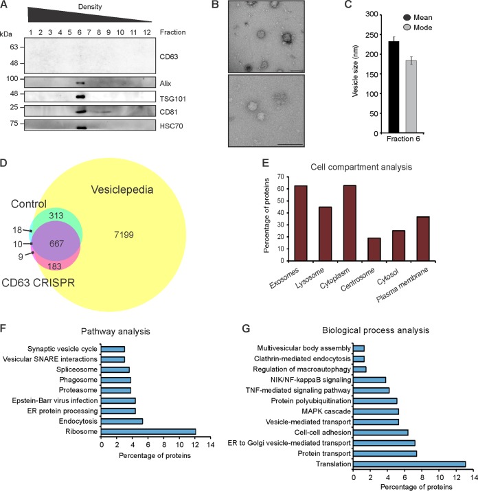
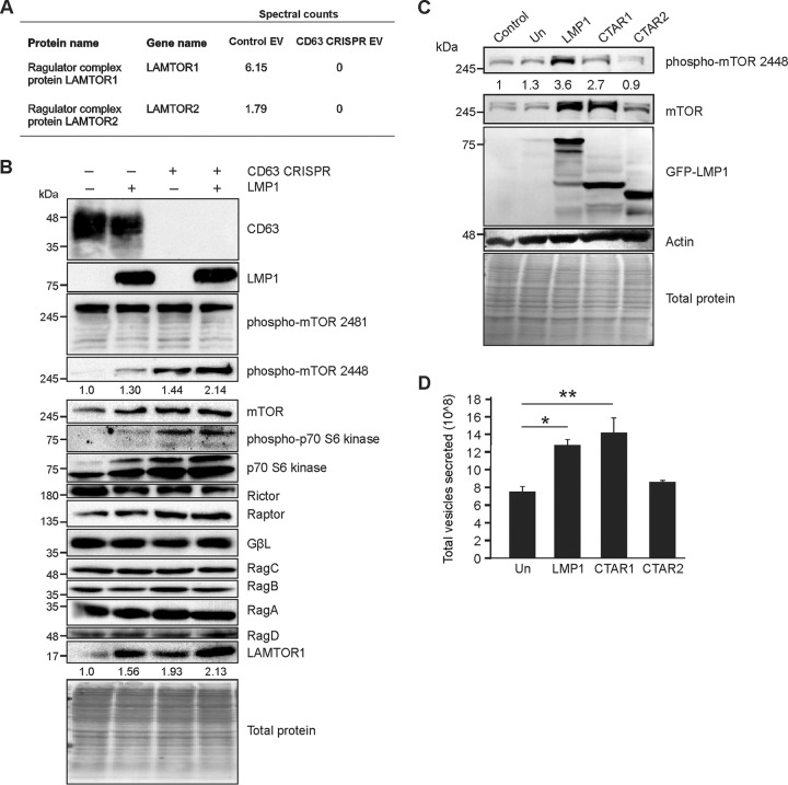
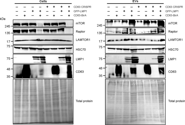
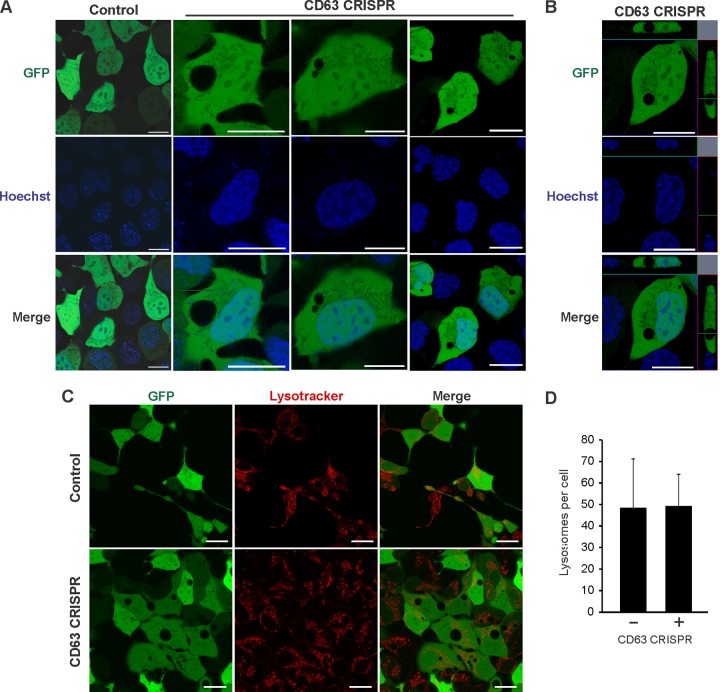
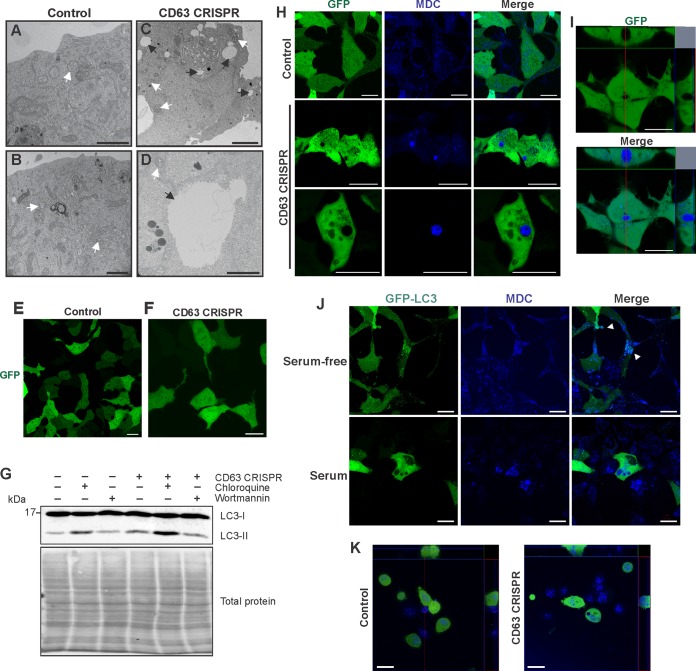
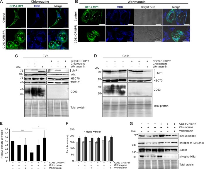
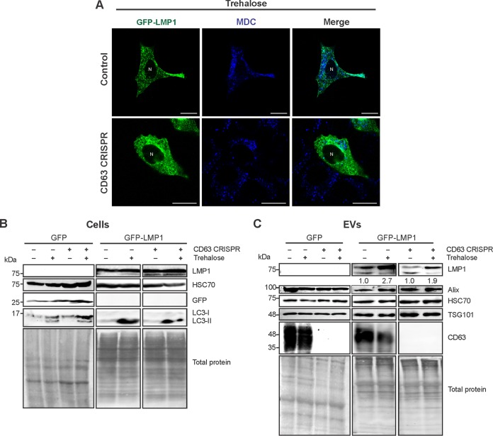
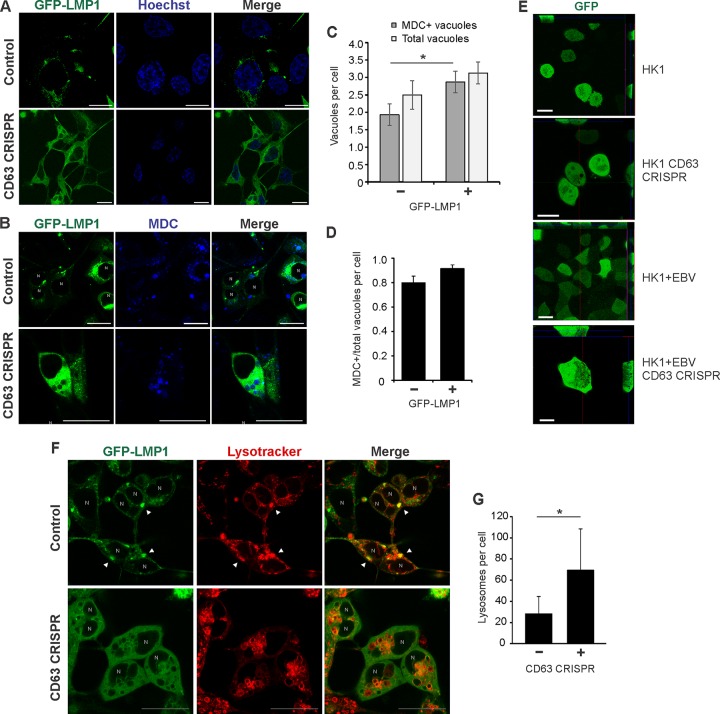
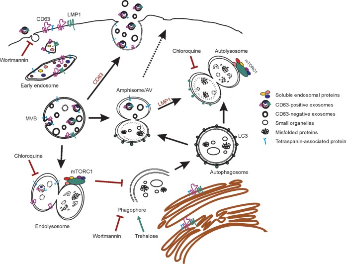
The tetraspanin protein CD63 has been recently described as a key factor in extracellular vesicle (EV) production and endosomal cargo sorting. In the context of Epstein-Barr virus (EBV) infection, CD63 is required for the efficient packaging of the major viral oncoprotein latent membrane protein 1 (LMP1) into exosomes and other EV populations and acts as a negative regulator of LMP1 intracellular signaling. Accumulating evidence has also pointed to intersections of the endosomal and autophagy pathways in maintaining cellular secretory processes and as sites for viral assembly and replication. Indeed, LMP1 can activate the mammalian target of rapamycin (mTOR) pathway to suppress host cell autophagy and facilitate cell growth and proliferation. Despite the growing recognition of cross talk between endosomes and autophagosomes and its relevance to viral infection, little is understood about the molecular mechanisms governing endosomal and autophagy convergence. Here, we demonstrate that CD63-dependent vesicle protein secretion directly opposes intracellular signaling activation downstream of LMP1, including mTOR-associated proteins. Conversely, disruption of normal autolysosomal processes increases LMP1 secretion and dampens signal transduction by the viral protein. Increases in mTOR activation following CD63 knockout are coincident with the development of serum-dependent autophagic vacuoles that are acidified in the presence of high LMP1 levels. Altogether, these findings suggest a key role of CD63 in regulating the interactions between endosomal and autophagy processes and limiting cellular signaling activity in both noninfected and virally infected cells.IMPORTANCE The close connection between extracellular vesicles and viruses is becoming rapidly and more widely appreciated. EBV, a human gamma herpesvirus that contributes to the progression of a multitude of lymphomas and carcinomas in immunocompromised or genetically susceptible populations, packages its major oncoprotein, LMP1, into vesicles for secretion. We have recently described a role of the host cell protein CD63 in regulating intracellular signaling of the viral oncoprotein by shuttling LMP1 into exosomes. Here, we provide strong evidence of the utility of CD63-dependent EVs in regulating global intracellular signaling, including mTOR activation by LMP1. We also demonstrate a key role of CD63 in coordinating endosomal and autophagic processes to regulate LMP1 levels within the cell. Overall, this study offers new insights into the complex intersection of cellular secretory and degradative mechanisms and the implications of these processes in viral replication.

Keywords: Epstein-Barr virus; amphisomes; exosomes; extracellular vesicles; herpesvirus; herpesviruses; multivesicular bodies.


Figures

FIG 1
FIG 1
CD63 is required for proper sorting of protein cargo into small extracellular vesicles. Small EVs derived from HEK293 control or CD63 CRISPR cells were purified by iodixanol density gradient ultracentrifugation for mass spectrometry analysis. (A) The presence of EVs in the fractions was determined by immunoblot analysis for common EV markers. A representative gradient of EVs purified from CD63 CRISPR cells is shown, with equal volumes loaded. The presence of EVs in fraction six of the gradient was confirmed by electron microscopy (B) and nanoparticle tracking analysis (C). (D) Overlap of proteins found in control or CD63 CRISPR cell-derived EVs identified by mass spectrometry compared to the Vesiclepedia database of EV proteins. (E) FunRich analysis of cellular compartments enriched in all proteins identified. Analysis of pathways (F) and biological processes (G) enriched in the data set of proteins decreased by 2-fold or greater in CD63 CRISPR EVs. Size bar, 250 nm.
FIG 2
FIG 2
CD63 negatively regulates mTORC1 signaling by Epstein-Barr virus LMP1. (A) LC-MS/MS spectral count quantification of LAMTOR proteins secreted in EVs from control and CD63 CRISPR cells. (B) HEK293 control or CD63 CRISPR cells were grown in serum-free medium and transfected with GFP or GFP-LMP1 for 24 h before cytoplasmic fractions were isolated, lysed, and analyzed by immunoblot analysis, with equal protein mass loaded. (C) Wild type (LMP1) and LMP1 C-terminal mutants containing the CTAR1 domain but lacking CTAR2 (CTAR1) or containing the CTAR2 domain but lacking CTAR1 (CTAR2) were induced by doxycycline in HK1 cells. Cell lysates were analyzed by immunoblotting for mTOR activation. All blots are representative images from 3 independent experiments. Quantitation of bands is based on averages from all experiments. (D) EVs were collected from HK1 cells following LMP1 mutant induction, and total vesicles were quantitated by nanoparticle tracking analysis. Un, uninduced. **, P < 0.01; *, P < 0.05.
FIG 3
FIG 3
Rescue of CD63 increases vesicle secretion of LMP1 and mTOR-associated proteins. GFP-LMP1 and fused CD63-BirA proteins were transfected into HEK293 control or CD63 CRISPR cells. Cell lysates and EVs were collected for immunoblot analysis of mTOR-associated protein levels 24 h after transfection. All blots are representative images from independent experiments.
FIG 4
FIG 4
CD63 CRISPR cells develop large nonlysosomal cellular compartments. (A) HEK293 control or CD63 CRISPR cells were transfected with GFP to visualize large cellular structures by confocal microscopy. Hoechst stain was used to identify nuclear compartments. (B) Orthogonal projection of a representative CD63 CRISPR cell. (C) Cells were stained by LysoTracker to examine acidified compartments within cells. (D) Quantification of total LysoTracker-positive compartments in cells (n = 20 cells). Size bars, 20 μm.
FIG 5
FIG 5
CD63 knockout induces the development of large autophagic vacuoles. HEK293 control (A and B) or CD63 CRISPR (C and D) cells were fixed and stained for transmission electron microscopy to visualize large intracellular vacuoles. Black arrows denote vacuoles, and white arrows denote MVBs. Size bar, 1 μm. HEK293 control (E) or CD63 CRISPR (F) cells were transfected with GFP following serum starvation for 24 h. (G) Cells were treated with chloroquine or wortmannin under serum-deprived conditions for 2 h. LC3 levels were measured by immunoblot analysis. Blots are representative of three independent experiments. (H) Cells were transfected with GFP and stained with mono-dansylcadaverine (MDC) to examine the morphology and localization of autophagic vacuoles. (I) Orthogonal sections of CD63 CRISPR cells stained with MDC. (J) HEK293 CD63 CRISPR cells were transfected with GFP-LC3 and grown under full-serum or serum-free conditions before staining with MDC to visualize vacuoles by confocal microscopy. (K) Nasopharyngeal carcinoma cells (HK1) and an HK1 CD63 CRISPR derivative cell line were transfected with GFP and stained with MDC to visualize autophagic vacuoles. Size bars, 20 μm.
FIG 6
FIG 6
Extracellular vesicle secretion and autophagic processes regulate LMP1-mediated intracellular signaling. Confocal microscopy of GFP-LMP1-transfected control or CD63 CRISPR cells following chloroquine (A) and wortmannin (B) treatment. Vacuoles were stained with MDC before live-cell imaging. Size bars, 20 μm. N, nuclear compartments. (C) Immunoblot analysis of EVs derived from HEK293 control or CD63 CRISPR cells following transfection of GFP-LMP1 and treatment with chloroquine or wortmannin for 24 h, with equal volumes loaded. (D) Immunoblots of corresponding cell lysates, with equal protein mass loaded. Nanoparticle tracking analysis of the quantity (E) and size (F) of EVs from cells following treatment with autophagy inhibitors. (G) Immunoblot analysis of cytoplasmic cellular fractions of cells transfected with GFP-LMP1, with equal protein mass loaded. Blots are representative images from repeated independent experiments. ***, P < 0.001; *, P < 0.05.
FIG 7
FIG 7
Autophagy activation by trehalose drives CD63-independent LMP1 release. (A) Confocal microscopy of GFP-LMP1-transfected control or CD63 CRISPR cells following trehalose treatment. Vacuoles were stained with MDC before live-cell imaging. Size bars, 20 μm. N, nuclear compartments. (B) Immunoblot analysis of cytoplasmic lysates from HEK293 control or CD63 CRISPR cells following transfection of GFP or GFP-LMP1 and treatment with trehalose for 24 h, with equal protein mass loaded. (C) Immunoblots of corresponding cell-derived EVs, with equal volumes loaded. Blots are representative images from repeated independent experiments. Quantitation of LMP1 secreted following trehalose treatment was normalized to parental cell secretion over multiple experiments.
FIG 8
FIG 8
LMP1 promotes acidification of CD63 knockout-induced autophagic vacuoles. HEK293 control and CD63 CRISPR cells were transfected with GFP-LMP1 and stained with Hoechst (A) and MDC (B). (C) MDC-positive and total vacuoles were quantified for each cell (n = 30 cells). (D) Proportion of MDC-positive over total vacuoles per CD63 CRISPR cell. (E) HK1 control and CD63 CRISPR cells were transfected with GFP to visualize vacuoles. EBV-infected HK1 (HK1 + EBV) control cells and cells containing CD63 CRISPR were treated similarly to visualize autophagic vacuoles. (F) Cells were transfected with GFP-LMP1 and stained with LysoTracker. Arrowheads represent colocalization of LMP1 with lysosomal compartments. (G) LysoTracker-positive compartments quantitated for each cell (n = 20 cells). N, nuclear compartments. *, P < 0.05.
FIG 9
FIG 9
Proposed model of CD63-mediated intersection between endosomal and autophagic processes. Endosomal intraluminal vesicles (ILVs) are dependent in part upon CD63 for secretion as exosomes into the extracellular space. Alternatively, multivesicular bodies (MVBs) carrying ILVs can fuse to lysosomes for degradation, where mTORC1 is activated. The autophagy pathway also converges on endosomal processes. Once autophagosomes mature from phagophores, they may fuse with MVBs to form amphisomes or autophagic vacuoles (AVs). AVs may also traffic to the plasma membrane to release EVs. Treatment of cells with chloroquine or wortmannin increases Epstein-Barr virus LMP1 secretion by inhibiting lysosomal acidification or phagophore formation, respectively, and results in decreased mTORC1 activation. Wortmannin also blocks EV uptake. Activation of autophagy by trehalose drives secretion of CD63-independent vesicles containing LMP1. Depletion of CD63 results in decreased EV secretion, likely shunting MVBs toward lysosomal or autophagosomal fusion. As a result, mTORC1 activation is increased and AVs accumulate in cells. Introduction of high levels of LMP1 drives late stages of autophagy and results in the acidification of AVs.

References

    1. Meckes DG, Shair KH, Marquitz AR, Kung CP, Edwards RH, Raab-Traub N. 2010. Human tumor virus utilizes exosomes for intercellular communication. Proc Natl Acad Sci U S A 107:20370–20375. doi: 10.1073/pnas.1014194107. - DOI - PMC - PubMed
    1. Meckes DG, Raab-Traub N. 2011. Microvesicles and viral infection. J Virol 85:12844–12854. doi: 10.1128/JVI.05853-11. - DOI - PMC - PubMed
    1. Meckes DG. 2015. Exosomal communication goes viral. J Virol 89:5200–5203. doi: 10.1128/JVI.02470-14. - DOI - PMC - PubMed
    1. Pegtel DM, Cosmopoulos K, Thorley-Lawson DA, van Eijndhoven MA, Hopmans ES, Lindenberg JL, de Gruijl TD, Würdinger T, Middeldorp JM. 2010. Functional delivery of viral miRNAs via exosomes. Proc Natl Acad Sci U S A 107:6328–6333. doi: 10.1073/pnas.0914843107. - DOI - PMC - PubMed
    1. Gould SJ, Booth AM, Hildreth JE. 2003. The Trojan exosome hypothesis. Proc Natl Acad Sci U S A 100:10592–10597. doi: 10.1073/pnas.1831413100. - DOI - PMC - PubMed

Publication types

MeSH terms