. 2018 Feb 12;92(5):e01850-17.
doi: 10.1128/JVI.01850-17. Print 2018 Mar 1.

Structural Insight into a Human Neutralizing Antibody against Influenza Virus H7N9

Affiliations

Structural Insight into a Human Neutralizing Antibody against Influenza Virus H7N9

Cong Chen et al. J Virol. .

Abstract

Since its first emergence in East China in early 2013, many cases of avian influenza A H7N9 have been reported. The disease has extended to 22 provinces in mainland China and some surrounding areas. Strategies to combat viral infection are urgently needed. We previously isolated a human monoclonal antibody, HNIgGA6, that neutralized the H7N9 virus both in vitro and in vivo In this study, we determined the crystal structure of viral hemagglutinin (HA) globular head bound to the fragment antigen-binding region (Fab) of HNIgGA6. The crystal structure shows that the tip of the HNIgGA6 heavy-chain complementarity-determining region 3 (HCDR3) directly interposes into the receptor binding site (RBS) and mimics, in many respects, the interaction of the sialic acid receptor. Three residues at Y98, H183, and E190, which are critical to human cellular receptor binding, are also essential for HNIgGA6 recognition. Meanwhile, dual mutations at V186G and L226Q in RBS were able to disrupt viral HA1 binding with the antibody. Our study provides a better understanding of the mechanism for protective antibody recognition and a sound foundation for the design of therapeutic drugs and vaccines against H7N9 influenza.IMPORTANCE Neutralization by antibody is one of the most important mechanisms for a host to defend against viral infections. Human-originated antibody HNIgGA6 was generated in response to the natural infectious H7N9 virus and showed potential for use in suppression of H7N9 infection, with possible therapeutic implications. The crystal structure of the HNIgGA6/HA1 complex provided new insight into the protective immune response to H7N9 virus in humans, as well as possibilities for the development of effective H7N9 pandemic vaccines and antiviral molecules.

Keywords: H7N9; neutralizing antibodies; structural biology.


Figures

FIG 1
FIG 1
High binding affinity of HA1 of the avian influenza A/H7N9 to human MAb HINgGA6. (a) ELISA was performed using purified viral HA1 and the purified antibody. The experiments were done in triplicate, and the results are presented as means with standard deviations. (b) BIAcore plots showing binding of viral HA1 to MAb HINgGA6. The MAb was immobilized on a CM5 chip, and a series of concentrations of HA1 were flowed over the immobilized MAb. Binding affinity (KD) values were calculated using a steady-state affinity model produced with BIAcore 3000 analysis software. (c) Sequence alignment of H7N9 (A/Anhui/1/2013) and avian H7N7 (A/pigeon/Wenzhou/559/2013) on HA1 domain. Dots indicate sequence identity with the H7N9 of strain A/Anhui/1/2013.
FIG 2
FIG 2
Crystal structure of the globular head complexed with Fab HNIgGA6. (a) Crystal structure of the globular head HA1 of H7N9-AH with HNIgGA6 Fab complex. The head is represented in magenta, the heavy chain is in yellow, and the light chain is in green. (b) Globular head and HINgGA6 Fab complex superimposed onto the A/Anhui/12013 HA trimer (PDB ID 4NJ0). HA1 is in purple and HA2 is in cyan on one of the three monomers. (c) Conservation surface of HA1. The score of residue conservation was performed according to the ConSurf server (http://consurf.tau.ac.il/2016/), based on the comparison of amino acid sequences of HAs. The full-length HA sequences were downloaded from Gisaid (http://platform.gisaid.org/). HINgGA6 contact residues are labeled and shown as sticks, with heavy chain in yellow and light chain in green. (d) Closer view of the HINgGA6 epitope. Residues are shown as magenta sticks. HA1 is shown in gray.
FIG 3
FIG 3
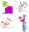
HINgGA6 binds to the HA receptor binding site using receptor mimicry. (a) Closeup view of the interaction between HNIgGA6 and H7 HA1. HA1 is in magenta and heavy chain is in yellow. Human receptor analog LSTc is pictured as orange sticks. (b and c) The HCDR3 of HNIgGA6 (colored in yellow and magenta) is inserted into the RBS of HA1 (silver) and binds in the same position as the LSTc (PDB ID 4N60). Hydrogen bonds are presented as dashed lines. (d) Three major modes of recognition by neutralizing antibodies against H7 HA. Two solved HA and antibody complex structures were docked onto monomeric A/Anhui/1/2013 H7 HA, presented in surface and colored in light blue. Antibody CT149 (PDB ID 4R8W) recognized residues in the stem of HA, and H7.167 (PDB ID 5F45) recognized residues adjacent to the RBS of HA, while HNIgGA6 binds to the RBS region.
FIG 4
FIG 4
Key residues involved in both antibody and receptor binding to HA. (a) Representation of amino acids in HA1 that share the interaction between HNIgGA6/HA and LSTc/HA. Key residues are highlighted in sticks. Binding of HNIgGA6 to HA mutant protein measured by ELISA. (b) Mutated HA proteins were expressed in HeLa cells and detected using IFA.
FIG 5
FIG 5
Involvement of V186 and L226 in antibody recognition. (a) Binding of HA mutants (V186G and L226Q) to MAb HNIgGA6 was measured by ELISA. (b) Dual mutations at V186G and L226Q resulted in loss of binding for HA to the antibody. (c) Structural basis for V186 and L226 in antibody recognition. Left, V186 and L226 create a favorable environment for HNIgGA6 binding. Right, the proposed model for 186G and 226Q in antibody recognition is displayed. The mutant residues are produced by the PYMOL program. The heavy chain (yellow) and HA1 (magenta and gray) are shown in cartoons, and residues are shown as sticks. The 130-loop, 190-helix, and 220-loop are colored in magenta. Potential hydrogen bonds are represented by dashed lines, with distances indicated.

References

    1. Garten RJ, Davis CT, Russell CA, Shu B, Lindstrom S, Balish A, Sessions WM, Xu X, Skepner E, Deyde V, Okomo-Adhiambo M, Gubareva L, Barnes J, Smith CB, Emery SL, Hillman MJ, Rivailler P, Smagala J, de Graaf M, Burke DF, Fouchier RA, Pappas C, Alpuche-Aranda CM, Lopez-Gatell H, Olivera H, Lopez I, Myers CA, Faix D, Blair PJ, Yu C, Keene KM, Dotson PD Jr, Boxrud D, Sambol AR, Abid SH, St George K, Bannerman T, Moore AL, Stringer DJ, Blevins P, Demmler-Harrison GJ, Ginsberg M, Kriner P, Waterman S, Smole S, Guevara HF, Belongia EA, Clark PA, Beatrice ST, Donis R, Katz J, Finelli L, Bridges CB, Shaw M, Jernigan DB, Uyeki TM, Smith DJ, Klimov AI, Cox NJ. 2009. Antigenic and genetic characteristics of swine-origin 2009 A(H1N1) influenza viruses circulating in humans. Science 325:197–201. doi:10.1126/science.1176225. - DOI - PMC - PubMed
    1. Kawaoka Y, Bean WJ, Webster RG. 1989. Evolution of the hemagglutinin of equine H3 influenza viruses. Virology 169:283–292. doi:10.1016/0042-6822(89)90153-0. - DOI - PubMed
    1. Palese P. 2004. Influenza: old and new threats. Nat Med 10:S82–S87. doi:10.1038/nm1141. - DOI - PubMed
    1. Scholtissek C, Rohde W, Von Hoyningen V, Rott R. 1978. On the origin of the human influenza virus subtypes H2N2 and H3N2. Virology 87:13–20. doi:10.1016/0042-6822(78)90153-8. - DOI - PubMed
    1. Peiris M, Yuen KY, Leung CW, Chan KH, Ip PL, Lai RW, Orr WK, Shortridge KF. 1999. Human infection with influenza H9N2. Lancet 354:916–917. doi:10.1016/S0140-6736(99)03311-5. - DOI - PubMed

Publication types

MeSH terms