. 2018 Jan 27;80(1):116-124.
doi: 10.1292/jvms.17-0569. Epub 2017 Dec 6.

Annexin A1 is a novel target gene of gonadotropin-releasing hormone in LβT2 gonadotrope cells

Affiliations

Annexin A1 is a novel target gene of gonadotropin-releasing hormone in LβT2 gonadotrope cells

Numfa Fungbun et al. J Vet Med Sci. .

Abstract

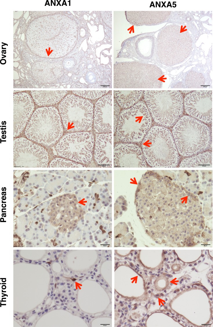
Gonadotropin-releasing hormone (GnRH) regulates gonadotropin secretion. We previously demonstrated that the expression of annexin A5 (ANXA5) is stimulated by GnRH in gonadotropes and has a significant role in gonadotropin secretion. It is therefore of interest to know whether other members of the ANXA family, which consists of twelve structurally related members, are also regulated by GnRH. Therefore, the expression of all annexins was examined in LβT2 gonadotrope cells. ANXA4, A5, A6, A7 and A11 were detected in LβT2 cells. The expression of ANXA5 and A1 mRNA was stimulated by a GnRH agonist. An increase in ANXA1 protein by this agonist was demonstrated by western blotting. Immunohistochemistry showed that ANXA1 was present in the nucleus and to a lesser extent in the cytoplasm of some rat pituitary cells. The GnRH agonist induced translocation of ANXA1 to the periphery of LβT2 cells. The presence of ANXA1 in gonadotropes and its increase upon GnRH agonist treatment were confirmed in a primary pituitary cell culture. ANXA1 expression was also demonstrated in the ovary, the testis, the thyroid gland and the pancreas in a different manner to that of ANXA5. These data suggest that ANXA1 is a novel GnRH target gene in gonadotropes. ANXA1 also may be a target of local GnRH in peripheral tissues and may have a different role than that of ANXA5.

Keywords: GnRH; annexin A1; annexin A5; cell biology; gonadotrope.


Figures

Fig. 1.
Fig. 1.
Rate of annexin mRNA expression in intact LβT2 cells. RNA was obtained from LβT2 cells and was used to determine the mRNA expression of each annexin gene using real time RT-PCR with specific primers. ND: not detectable. Each annexin expression rate is expressed as a ratio to the level of ANXA5 mRNA.
Fig. 2.
Fig. 2.
Effects of GnRH agonist treatment on annexin mRNA expression. The mRNA expression of ANXA1, A4, A5, A6, A7 and A11 in LβT2 cells was examined using real time RT-PCR after 12 hr of incubation with 10−7 M of the GnRH agonist (GnRHa). Asterisks indicate a significant difference from the respective control value, P<0.05.
Fig. 3.
Fig. 3.
Changes in the rate of ANXA1 and A5 mRNA expression over time in LβT2 cells after GnRH agonist stimulation. The GnRH agonist (10−7 M) was administered to LβT2 cells and the cells were harvested after 0, 1, 3 and 9 hr of incubation. ANXA1 and A5 mRNA was then analyzed using real time RT-PCR. Asterisks indicate a significant difference from the 0 hr value of each group, P<0.01.
Fig. 4.
Fig. 4.
ANXA1 protein expression after GnRH agonist stimulation. ANXA1 protein expression was estimated by western blotting after GnRH agonist administration. LβT2 cells were challenged with 10−7 M of the GnRH agonist for 3 hr and cell lysates were analyzed using SDS-PAGE and western blotting with an anti-ANXA1 antibody. (A) Immunostaining image after western blotting. Lanes 1–4 are control incubations and lanes 5–8 are GnRH agonist-treated samples. β-actin was used as an internal control. Images were cropped and the original membranes are shown in Supplementary figure. (B) The intensity of each band was measured by densitometry. Intensities were normalized with that of the internal control (β-actin). Values are the mean of four samples with the standard error. An asterisk indicates a significant difference, P<0.05.
Fig. 5.
Fig. 5.
Immunohistochemical and immunofluorescence analysis of annexin expression. A: ANXA1 and A5 expression in pituitary tissues of female rats was analyzed using immunohistochemistry. B: LβT2 cells were grown on a coverslip and challenged with the GnRH agonist (10−9 M) at time 0, then ANXA1 expression over time was analyzed with an anti-ANXA1 antibody using immunofluorescence. ANXA1 is shown with green fluorescence. DAPI was used to stain the nucleus (blue). Red arrows indicate accumulation of ANXA1 in immunoreactive puncta. Yellow arrows indicate ANXA1 accumulation at the periphery of cells. C: ANXA1 accumulation (yellow arrow) to the periphery of LβT2 cells (b) shown with a differential interference contrast (dic) image (a). Actin is shown in red from phalloidin staining. ANXA1 is shown with green fluorescence. DAPI was used to stain the nucleus (blue). D: The coexistence of ANXA1 and the LHβ subunit was shown with double staining immunohistochemistry. Cells were stained with anti-LHβ (red) and anti-ANXA1 (green). Nuclei were stained with DAPI (blue). a: negative control for ANXA1. The cells were stained without anti-ANXA1. b: Cells were stained before exposure to the GnRH agonist. c: The cell after 48 hr of incubation with GnRH agonist (10−9 M) was double stained. Yellow arrow indicates ANXA1 in the cytoplasm.
Fig. 6.
Fig. 6.
Immunohistochemical analysis of ANXA1 and A5 expression in endocrine organs. ANXA1 and A5 expression in rat ovary, testis, pancreas and thyroid tissues was analyzed using immunohistochemistry. Red arrows represent a typical positive reaction in each tissue.

References

    1. Buckingham J. C.1996. Fifteenth Gaddum Memorial Lecture December 1994. Stress and the neuroendocrine-immune axis: the pivotal role of glucocorticoids and lipocortin 1. Br. J. Pharmacol. 118: 1–19. doi: 10.1111/j.1476-5381.1996.tb15360.x - DOI - PMC - PubMed
    1. Buckingham J. C., Flower R. J.1997. Lipocortin 1: a second messenger of glucocorticoid action in the hypothalamo-pituitary-adrenocortical axis. Mol. Med. Today 3: 296–302. doi: 10.1016/S1357-4310(97)88908-3 - DOI - PubMed
    1. Cirino G., Peers S. H., Flower R. J., Browning J. L., Pepinsky R. B.1989. Human recombinant lipocortin 1 has acute local anti-inflammatory properties in the rat paw edema test. Proc. Natl. Acad. Sci. U.S.A. 86: 3428–3432. doi: 10.1073/pnas.86.9.3428 - DOI - PMC - PubMed
    1. Crompton M. R., Moss S. E., Crumpton M. J.1988. Diversity in the lipocortin/calpactin family. Cell 55: 1–3. doi: 10.1016/0092-8674(88)90002-5 - DOI - PubMed
    1. De Loof A., Lindemans M., Liu F., De Groef B., Schoofs L.2012. Endocrine archeology: do insects retain ancestrally inherited counterparts of the vertebrate releasing hormones GnRH, GHRH, TRH, and CRF? Gen. Comp. Endocrinol. 177: 18–27. doi: 10.1016/j.ygcen.2012.02.002 - DOI - PubMed

MeSH terms