. 2017 Dec 6;7(1):17036.
doi: 10.1038/s41598-017-15986-2.

In vivo evidence for homo- and heterodimeric interactions of Arabidopsis thaliana dehydrins AtCOR47, AtERD10, and AtRAB18

Affiliations

In vivo evidence for homo- and heterodimeric interactions of Arabidopsis thaliana dehydrins AtCOR47, AtERD10, and AtRAB18

Itzell E Hernández-Sánchez et al. Sci Rep. .

Abstract

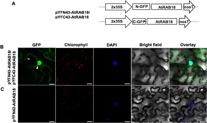
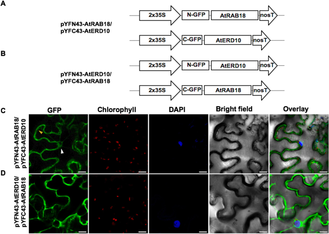
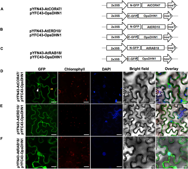
Dehydrins (DHNs) are intrinsically disordered proteins that play central roles in plant abiotic stress responses; however, how they work remains unclear. Herein, we report the in planta subcellular localization of Arabidopsis thaliana DHNs AtCOR47, AtERD10, and AtRAB18 through GFP translational fusions. To explore the dimerization ability of the Arabidopsis acidic DHNs AtCOR47 and AtERD10, we conducted an in planta DHN binding assay using the Bimolecular Fluorescence Complementation (BiFC) technique. Our analyses revealed homodimeric interactions for AtCOR47 and AtERD10; interestingly, heterodimeric associations also occurred with these DHNs, and these interactions were observed in the cytosol of tobacco cells. Furthermore, we evaluated whether Arabidopsis basic DHNs, such as AtRAB18, could also interact with itself and/or with AtCOR47 and AtERD10 in the BiFC system. Our data revealed homodimeric RAB18 complexes in the nucleus and cytosol, while heterodimeric associations between AtRAB18 and acidic DHNs occurred only in the cytosol. Finally, we demonstrated the presence of heterodimeric complexes among Arabidopsis AtCOR47, AtERD10, and AtRAB18 DHNs with their acidic ortholog the OpsDHN1 from Opuntia streptacantha; these heterodimeric interactions showed different subcellular distributions. Our results guide DHN research toward a new scenario where DHN/DHN oligomerization could be explored as a part of their molecular mechanism.


Conflict of interest statement

The authors declare that they have no competing interests.

Figures

Figure 1
Figure 1
Determination of the AtCOR47 homodimers using the BiFC assay. (A) Schematic diagram of pYFN43-AtCOR47 and pYFC43-AtCOR47 BiFC vectors. (B) Confocal images of AtCOR47/AtCOR47 interaction. (C) Auto-fluorescence test in the BiFC system of the pYFN43-AtCOR47 construct transformed alone. From left to right: GFP, chlorophyll, DAPI, bright field, and overlay channels, white arrow indicates the nuclei and yellow arrow indicates the cytosol. The scale bar corresponds to 23 μm.
Figure 2
Figure 2
Visualization of the AtERD10 homodimers using the BiFC assay. (A) Schematic representation of BiFC vectors containing the AtERD10 protein fused to N- or C-terminus of GFP. (B) Fluorescence analysis of AtERD10/AtERD10 interaction. (C) Analysis for no fluorescent auto-activation of the pYFN43-AtERD10 construct. From left to right: GFP, chlorophyll, DAPI, bright field and overlay channels, white arrow indicates the nuclei and yellow indicates the cytosol. The scale bar corresponds to 23 μm.
Figure 3
Figure 3
Detection of the basic AtRAB18 homodimer in BiFC assay. (A) Schematic representation of BiFC vectors containing the AtRAB18 protein fused to pYFN43 or pYFC43 vectors. (B) Fluorescence analysis of AtRAB18/AtRAB18 interaction. (C) Auto-fluorescence test of the pYFN43-AtRAB18 vector. From left to right: GFP, chlorophyll, DAPI, bright field and overlay panels. The white arrow indicates the nuclei and yellow arrow indicates the cytosol. The scale bar corresponds to 23 μm.
Figure 4
Figure 4
The AtCOR47 and AtERD10 DHNs are able to heterodimerize in BiFC assay. Schematic representation of the (A) pYFN43/pYFC43-AtCOR47 and (B) pYFN43/pYFC43-AtERD10 BiFC constructs. Confocal images of: (C) AtCOR47/AtERD10 and (D) AtERD10/AtCOR47 BiFC interactions. From left to right: GFP, chlorophyll, DAPI, bright field and overlay channels, white arrow indicates the nuclei and yellow arrow indicates the cytosol. The scale bar corresponds to 23 μm.
Figure 5
Figure 5
Heterodimerization between the Arabidopsis dehydrins: AtCOR47 and AtRAB18 in BiFC assay. (A) Schematic representation of the pYFN43-AtRAB18/pYFC43-AtCOR47 and (B) pYFN43-AtCOR47/pYFC43-AtRAB18 constructs. Fluorescent analyses of (C) AtRAB18/AtCOR47 and (D) AtCOR47/AtRAB18 interactions. From left to right: GFP, chlorophyll, DAPI, bright field and overlay channels, white arrow indicates the nuclei and yellow arrow targets the cytosol. The scale bar corresponds to 23 μm.
Figure 6
Figure 6
Identification of the AtERD10/AtRAB18 heterodimer interaction using the BiFC assay. Schematic illustration of the BiFC constructs containing the AtERD10 and AtRAB18 proteins. (A) The pYFN43-AtRAB18/pYFC43-AtERD10 constructs. (B) The pYFN43-AtERD10/pYFC43-AtRAB18 vectors. (C,D) Fluorescence visualization of AtRAB18/AtERD10 heterodimer formation. From left to right: GFP, chlorophyll, DAPI, bright field and overlay channels, white arrow indicates the nuclei and yellow arrows target the cytosol. The scale bar corresponds to 23 μm.
Figure 7
Figure 7
BiFC analysis of heterodimeric interaction among the Arabidopsis dehydrins: AtCOR47, AtERD10 and AtRAB18 with its orthologue OpsDHN1. Diagrammatic illustration of the BiFC constructs AtCOR47, AtERD10, AtRAB18 and OpsDHN1. (A) The pYFN43-AtCOR47/pYFC43-OpsDHN1 vectors. (B) The pYFN43-AtERD10/pYFC43-OpsDHN1 vectors. (C) The pYFN43-AtRAB18/pYFC43-OpsDHN1 vectors. Confocal interaction analysis of: (D) AtCOR47/OpsDHN1. (E) AtERD10/OpsDHN1. (F) AtRAB18/OpsDHN1. From left to right: GFP, chlorophyll, DAPI, bright field and overlay channels, white arrow indicates the nuclei and yellow arrows target the cytosol. The scale bar corresponds to 23 μm.
Figure 8
Figure 8
Analysis of interactions between OpsDHN1ΔHis version and AtCOR47 DHNs in BiFC system. Schematic depiction of: (A) pYFN43-OpsDHN1ΔHis/pYFC43AtCOR47. (B) pYFN43-AtCOR47/pYFC43-OpsDHN1ΔHis. (C,D) Confocal analysis of: OpsDHN1ΔHis/AtCOR47 interaction or its swapped version. From left to right: GFP, chlorophyll, DAPI, bright field and overlay channels, white arrow indicates the nuclei and yellow arrows target the cytosol. The scale bar corresponds to 23 μm.
Figure 9
Figure 9
BiFC analysis of OpsDHN1ΔHis version and AtERD10 DHNs. Graphic representation of assayed constructs (A) pYFN43-OpsDHN1ΔHis/pYFC43-AtERD10 and (B) pYFN43-AtERD10/pYFC43-OpsDHN1ΔHis. Confocal pictures of: (C) OpsDHN1ΔHis/AtERD10 and (D) AtERD10/OpsDHN1ΔHis interactions. From left to right: GFP, chlorophyll, DAPI, bright field and overlay channels, white arrow indicates the nuclei and yellow arrows target the cytosol. The scale bar corresponds to 23 μm.
Figure 10
Figure 10
Interaction assay of OpsDHN1ΔHis version and AtRAB18 DHNs in BiFC system. A simplified representation of BiFC constructs. (A) pYFN43-OpsDHN1ΔHis/pYFC43-AtRAB18 and (B) pYFN43-AtRAB18/pYFC43-OpsDHN1ΔHis. Confocal pictures of transient co-transformed vectors. (C) OpsDHN1ΔHis/AtRAB18 and (D) AtRAB18/OpsDHN1ΔHis. From left to right: GFP, chlorophyll, DAPI, bright field and overlay channels, white arrow indicates the nuclei and yellow arrows target the cytosol. The scale bar corresponds to 23 μm.
Figure 11
Figure 11
Proposed model for DHN dimerization in plant cells. Representation of homo- and heteromeric interaction among Arabidopsis dehydrins, and its interaction with the Opuntia streptacantha OpsDHN1 in tobacco cells. According to their localization results, the interactions AtCOR47/AtCOR47, AtCOR47/OpsDHN1 AtCOR47/AtERD10 and AtERD10/AtERD10, AtERD10/OpsDHN1 proteins only occur in the cytosol and could be protecting proteins from damage caused by stress. In contrast, AtRAB18/AtRAB18 and AtRAB18/OpsDHN1 were observed in both the cytosol and nucleus and could be protecting proteins from stress damage in either subcellular sites or by interacting with nucleic acids. This figure was created using Somersault 18:24 images as templates (http://www.somersault1824.com/science-illustrations/).

References

    1. Ahuja I, de Vos RCH, Bones AM, Hall RD. Plant molecular stress responses face climate change. Trends Plant Sci. 2010;15:664–674. doi: 10.1016/j.tplants.2010.08.002. - DOI - PubMed
    1. Hanin M, Brini FB, Takeda TY, Masmoudi K. Plant dehydrins and stress tolerance: versatile proteins for complex mechanisms. Plant Signal Behav. 2011;6:1503–1509. doi: 10.4161/psb.6.10.17088. - DOI - PMC - PubMed
    1. Welin BV, Olson Å, Nylander M, Palva ET. Characterization and differential expression of dhn/lea/rab-like genes during cold acclimation and drought stress in Arabidopsis thaliana. Plant Mol. Biol. 1994;26:131–144. doi: 10.1007/BF00039526. - DOI - PubMed
    1. Dhanaraj AL, Slovin JP, Rowland LJ. Analysis of gene expression associated with cold acclimation in blueberry floral buds using expressed sequence tags. Plant Sci. 2004;166:863–872. doi: 10.1016/j.plantsci.2003.11.013. - DOI
    1. Arumingtyas EL, Savitri ES, Purwoningrahayu RD. Protein profiles and dehydrin accumulation in some soybean varieties (Glycine max L. Merr) in drought stress conditions. Am J Plant Sci. 2013;4:134–141. doi: 10.4236/ajps.2013.41018. - DOI

Publication types

Substances

LinkOut - more resources