. 2017 Dec 6;7(1):17095.
doi: 10.1038/s41598-017-16609-6.

Exosomes from antigen-pulsed dendritic cells induce stronger antigen-specific immune responses than microvesicles in vivo

Affiliations

Exosomes from antigen-pulsed dendritic cells induce stronger antigen-specific immune responses than microvesicles in vivo

Casper J E Wahlund et al. Sci Rep. .

Abstract

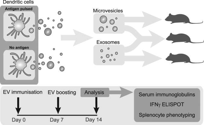
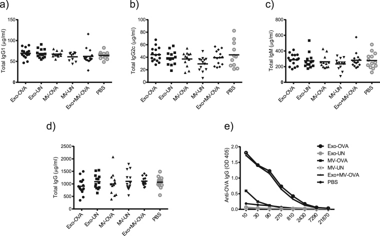
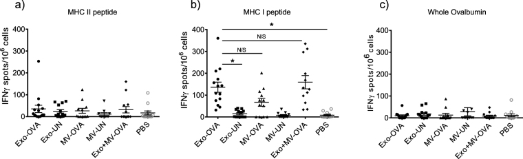
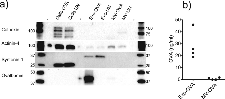
Extracellular vesicles (EV), including exosomes and microvesicles (MV), represent a rapidly expanding field of research with diagnostic and therapeutic applications. Although many aspects of EV function remain to be revealed and broad investigations are warranted, most published findings focus on only one vesicle category or a non-separated mix of EVs. In this paper, we investigated both MVs and exosomes from Ovalbumin (OVA)-pulsed dendritic cells for their immunostimulatory potential side-by-side in vivo. Only exosomes induced antigen-specific CD8+ T-cells, and were more efficient than MVs in eliciting antigen-specific IgG production. Further, mainly exosome-primed mouse splenocytes showed significant ex vivo interferon gamma production in response to antigen restimulation. Exosomes carried high levels of OVA, while OVA in MVs was barely detectable, which could explain the more potent antigen-specific response induced by exosomes. Moreover, exosomes induced increased germinal center B cell proportions, whereas MVs had no such effect. Immunisation with both vesicle types combined showed neither inhibitory nor synergistic effects. We conclude that DC-derived MVs and exosomes differ in their capacity to incorporate antigen and induce immune responses. The results are of importance for understanding the role of EVs in vivo, and for future design of vesicle-based immunotherapies and vaccines.


Conflict of interest statement

Gabrielsson declares a potential conflict of interest with a US patent on B-cell derived exosome-based immune therapy. No other authors declare any conflicts of interest.

Figures

Figure 1
Figure 1
Exosomes and microvesicles from OVA-pulsed dendritic cells (DC) display similar levels of immunorelevant markers and tetraspanins. Using polystyrene-latex coated with anti-MHC class II antibodies, vesicles corresponding to the same total protein amounts were captured and analysed by flow cytometry. Quantified expression levels in mean fluorescence intensity of MHC class I and II, the adhesion molecule CD54, costimulatory CD86, CD80 and CD40 as well as the tetraspanins CD9, CD63 and CD81. All levels are MFI ratio of each marker normalised to isotype controls, conducted on four batches of exosomes (Exo) and microvesicles (MV) from OVA-pulsed (OVA) or un-pulsed DCs (UN).
Figure 2
Figure 2
Microvesicles and exosomes have similar sizes and characteristics. Nanoparticle Tracking Analysis (NTA) showed overlapping size distributions of exosomes (a) and microvesicles (MV) (b), a mean diameter of 153 nm for exosomes and 170 nm for MVs (p = 0.026, c) and equal numbers of vesicles released per cell (d). Results are pooled of three independent experiments conducted on in total six separate preparations per vesicle type. Five 60 second recordings per sample were batch analysed. Transmission electron microscopy showed typical characteristics of exosomes (e) and similar, but generally larger sizes of MVs (f). Size bars (in e and f) are 200 nm per segment (black or white).
Figure 3
Figure 3
Experimental layout for in vivo analysis of immune responses to antigen-loaded extracellular vesicles. Exosomes and microvesicles from antigen-pulsed or non-pulsed bone marrow-derived dendritic cells were isolated, and injected at days zero and seven. On day 14, blood and spleens were analysed for serum immunoglobulins, spleen immune cell characterisation and interferon gamma (IFNγ) response to ex vivo antigenic restimulation.
Figure 4
Figure 4
OVA-loaded exosomes (Exo-OVA) but not microvesicles (MV-OVA) induce immune cell population changes in vivo. Exo-OVA, MV-OVA or a combination thereof was injected on day zero and day seven in C57Bl/6 mice, and the splenocytes were characterised by flow cytometry on day 14. Percentages of B220+ B cells (a), GL-7+CD95+ germinal center B cells (b), CD138+ plasma cells (c), CD3+B220- T cells (d), CD3+CD4<sup>−</sup> CD8+T cells (e) and OVA-specific CD8+T cells (f) are displayed. Control groups were PBS, non-OVA loaded (UN) vesicles and PBS alone. Results are pooled from at least three experiments. (n = 4–6 per group per experiment). *Indicates p < 0.05, **indicates p < 0.01 and ***indicates p < 0.001, one-way ANOVA test with Dunn’s multiple correction.
Figure 5
Figure 5
Exosomes induce higher antigen-specific IgG levels compared to microvesicles. Day 14 serum levels of mice immunised with exosomes (Exo-OVA), microvesicles (MV-OVA) or a mix of both vesicle types from 18 million OVA-pulsed dendritic cells was analysed for levels of total IgG1 (a), total IgG2c (b), total IgM (c), total IgG (d), and OVA-specific IgG (e). Results are pooled from three independent experiments (n = 4–7 per group per experiment). Control groups were non-antigen loaded (UN) vesicles and PBS. *Indicates p < 0.05, ***Indicates p < 0.001, one-way ANOVA test with Dunn’s multiple correction.
Figure 6
Figure 6
Splenocytes from all mice injected with OVA-loaded exosomes (Exo-OVA), and majority of those injected with microvesicles (MV-OVA) are responsive to antigenic restimulation. Splenocytes from mice immunised with either Exo-OVA, MV-OVA or a combination of the two were restimulated in an IFNγ ELISPOT. Numbers of IFNγ-producing units were counted after restimulation with the MHC class II-associated OVA323–339 peptide (a), the MHC class I-associated OVA peptide SIINFEKL (b), or with whole OVA protein (c). Control groups were immunised with vesicles without antigen (Exo-UN, MV-UN) or PBS. Results are pooled from at least three independent experiments (n = 4–7 per group per experiment). *Indicates p < 0.05 and ***Indicates p < 0.001, one-way ANOVA test with Dunn´s multiple correction was used.
Figure 7
Figure 7
Exosomes are enriched in endosomal protein and incorporated antigen compared to microvesicles. (a) Western blot analysis of dendritic cell (DC) lysates and DC-derived vesicles for (from top) endoplasmic reticulum-associated Calnexin, MV-associated Actinin-4, ALIX-interacting Syntenin-1, Ovalbumin. (b) ELISA analysis of surface-oriented levels of Ovalbumin on four separate batches of exosomes and microvesicles. Representative of two experiments (a) or pooled result of four experiments (b). For b, a two-tailed t-test (Kruskal-Wallis) was performed. Uncropped gel images are displayed in Supplementary Figure S3.

References

    1. Jia S, et al. Emerging technologies in extracellular vesicle-based molecular diagnostics. Expert review of molecular diagnostics. 2014;14:307–321. doi: 10.1586/14737159.2014.893828. - DOI - PubMed
    1. Raposo G, Stoorvogel W. Extracellular vesicles: exosomes, microvesicles, and friends. The Journal of cell biology. 2013;200:373–383. doi: 10.1083/jcb.201211138. - DOI - PMC - PubMed
    1. S. ELA, Mager I, Breakefield XO, Wood MJ. Extracellular vesicles: biology and emerging therapeutic opportunities. Nature reviews. Drug discovery. 2013;12:347–357. doi: 10.1038/nrd3978. - DOI - PubMed
    1. Ostrowski, M. et al. Rab27a and Rab27b control different steps of the exosome secretion pathway. Nature cell biology12, 19–30; sup pp 11–13, 10.1038/ncb2000 (2010). - PubMed
    1. Colombo M, et al. Analysis of ESCRT functions in exosome biogenesis, composition and secretion highlights the heterogeneity of extracellular vesicles. Journal of cell science. 2013;126:5553–5565. doi: 10.1242/jcs.128868. - DOI - PubMed

Publication types

MeSH terms