. 2017 Dec 6;8(1):1968.
doi: 10.1038/s41467-017-02126-7.

Galectin-3 impacts Cryptococcus neoformans infection through direct antifungal effects

Affiliations

Galectin-3 impacts Cryptococcus neoformans infection through direct antifungal effects

Fausto Almeida et al. Nat Commun. .

Abstract

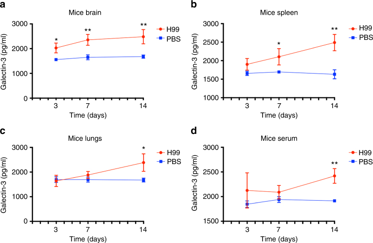
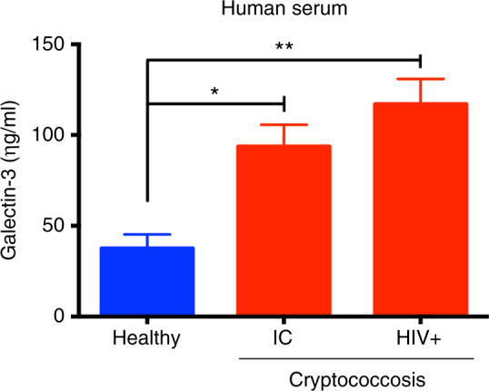
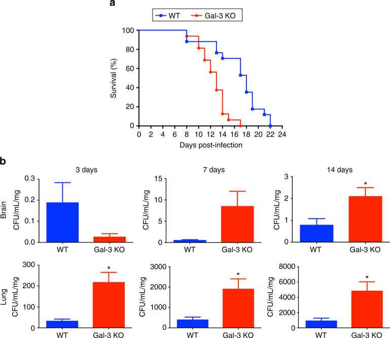
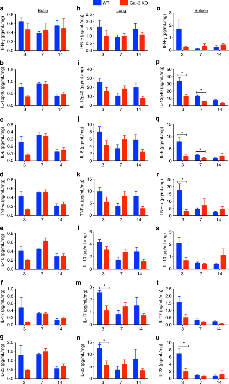
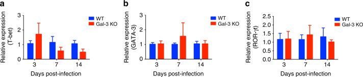
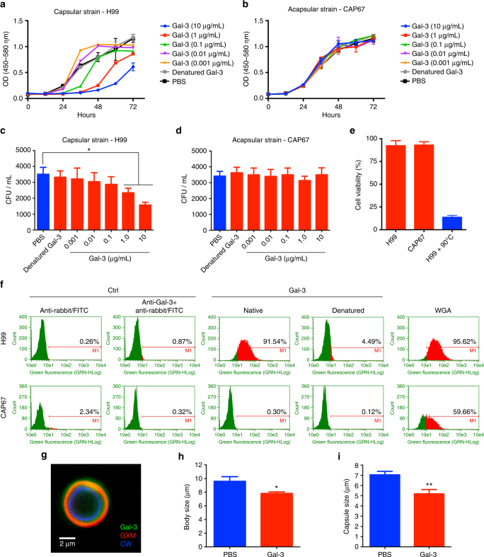
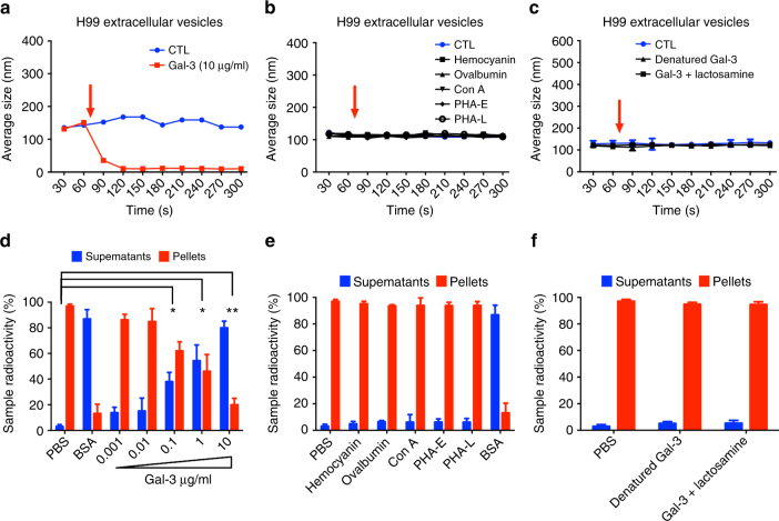
Cryptococcus neoformans is an encapsulated fungal pathogen that causes cryptococcosis, which is a major opportunistic infection in immunosuppressed individuals. Mammalian β-galactoside-binding protein Galectin-3 (Gal-3) modulates the host innate and adaptive immunity, and plays significant roles during microbial infections including some fungal diseases. Here we show that this protein plays a role also in C. neoformans infection. We find augmented Gal-3 serum levels in human and experimental infections, as well as in spleen, lung, and brain tissues of infected mice. Gal-3-deficient mice are more susceptible to cryptococcosis than WT animals, as demonstrated by the higher fungal burden and lower animal survival. In vitro experiments show that Gal-3 inhibits fungal growth and exerts a direct lytic effect on C. neoformans extracellular vesicles (EVs). Our results indicate a direct role for Gal-3 in antifungal immunity whereby this molecule affects the outcome of C. neoformans infection by inhibiting fungal growth and reducing EV stability, which in turn could benefit the host.


Conflict of interest statement

The authors declare no competing financial interests.

Figures

Fig. 1
Fig. 1
Upregulated Gal-3 levels in mice during experimental C. neoformans infection. C57BL/6 mice were intratracheally infected with H99 yeast cells (red line with circle) or PBS (blue line with square) and Gal-3 levels were verified in tissues and serum during the course of C. neoformans infection. On days 3, 7, and 14 after the fungal inoculation, samples collected of brain (a), spleen (b), lungs (c), and serum (d), were homogenized and assessed by ELISA regarding the Gal-3 concentration. Gal-3 levels were upregulated over time after infection with C. neoformans. Bars represent the mean ± SD of Gal-3 levels obtained from triplicate samples in groups of five animals. Statistically significant differences are denoted by asterisks (*p < 0.05, **p < 0.005, unpaired Student’s t-test)
Fig. 2
Fig. 2
Upregulated Gal-3 levels in humans during C. neoformans infection. Gal-3 levels in serum from healthy individuals (blue bar) and patients infected by C. neoformans (red bars) were assessed by ELISA. Gal-3 levels were higher in IC (immunocompetent) and HIV (human immunodeficiency virus) patients infected with C. neoformans when compared with healthy individuals. Bars represent the mean ± SD of Gal-3 levels obtained from triplicate samples. Statistically significant differences are denoted by asterisks (*p < 0.05, **p < 0.005, unpaired Student’s t-test)
Fig. 3
Fig. 3
Absence of Gal-3 leads to lower survival and increased fungal burden in experimental murine cryptococcosis. The survival rate (a) of Gal-3 KO mice (red line with circle) and WT mice (blue line with square) were verified after intratracheally infection with H99 yeast cells. b Colony-forming units (CFU) recovered from brain and lungs of WT (blue bars) and Gal-3 KO (red bars) mice were assessed after 3, 7, and 14 days after intratracheally infected with C. neoformans. The y-axis denotes CFU/ml/mg resulting from normalization of fungal burden to the weight of the organ fragment used to prepare the homogenate. Data are representative of three experiments, each performed with eight mice per group. The bars represent the mean ± SD of CFU obtained from triplicate samples in groups of eight animals. *p < 0.05, Student’s t-test, Gal-3 KO mice compared with WT mice
Fig. 4
Fig. 4
Levels of relevant cytokines in the brain, lung, and spleen of C. neoformans-infected WT and Gal-3 KO mice. WT (blue bars) and Gal-3 KO (red bars) mice were intratracheally infected with a 50 μl suspension containing 1×10⁶ yeast cells. Organ samples were weighed and homogenized. The levels of the following cytokines in the brains (ag), lungs (hn), and spleens (ou) (3, 7, and 14 days post-infection) of infected mice were determined by ELISA: IFN-γ, IL-12p40, IL-6, TNF-α, IL-10, IL-17, and IL-23. The results represent the mean ± SD of five mice per group, from a representative experiment of three assays. *p < 0.05, Student’s t-test, Gal-3 KO mice compared with WT mice
Fig. 5
Fig. 5
Levels of T-bet, GATA-3, and ROR-γt in the lungs of C. neoformans-infected WT and Gal-3 KO mice. WT (blue bars) and Gal-3 KO (red bars) mice were intratracheally infected with H99 yeast cells and samples of their lungs weighed and homogeneized. The levels of mRNA relative expression for T-bet (a), GATA-3 (b), and ROR-γt (c) in the lungs (3, 7, and 14 days post-infection) of infected mice were determined by real-time PCR, using the β-actin gene as control. The results represent the mean ± SD of five mice per group, from a representative experiment of three assays
Fig. 6
Fig. 6
Gal-3 interacts with the C. neoformans capsule and inhibits the fungal growth. Capsular strain H99 (a and c) and acapsular strain CAP67 (b and d) were incubated for 72 h at 37 °C with Gal-3 (0.001 to 10 μg/ml), and the optical density at 540 nm (OD540) measured every hour (a and b), or the number or colony-forming units (CFU) were counted for determination of cell densities (c and d). e H99 and CAP67 strains were incubated for 72 h at 37 °C with Gal-3 (10 μg/ml), and the frequency of viable cells assessed using propidium iodide staining. Heated fungal cells (90 °C for 10 min) were used as positive control (blue bar). H99 and CAP67 strains were incubated for 40 min at 4 °C with Gal-3 40 μg/ml. After that, both strains were washed with PBS and incubated for 45 min with anti-Gal-3 antibody. Then, washed again with PBS and incubated with anti-rabbit IgG-FITC antibody for 40 min at 4 °C. Labeled cells were acquired on a FACS Guava easyCyte, and the histogram represents the percentage of positive cells recognized by Gal-3 (f). Anti-rabbit IgG-FITC antibody associated or not with Gal-3 was used as control, and use of WGA lectin (30 μg/ml) as control of binding with capsule or cell wall (f). g H99 strain was cultured at 37 °C for 24 h and incubated with Gal-3. C. neoformans were stained for observation of cell wall (CW) with calcofluor white (blue), Gal-3 with anti-Gal-3 antibody (green), and capsule with anti-GXM (red). Body (h), and capsule (i) size of H99 strain cells were assessed by microscopic analysis after cultivation in the presence of Gal-3 10 μg/ml. Data are representative of three experiments. Statistically significant differences are denoted by asterisk (*p < 0.05, unpaired Student’s t-test)
Fig. 7
Fig. 7
Gal-3 disrupts C. neoformans extracellular vesicles. Temporal kinetics of vesicle disruption mediated by Gal-3 were measured by dynamic light scattering over ten 30 s to read the population average size (ac). The addition of Gal-3 10 μg initially leads to disruption (a). The same samples were read first without (blue line) and then with the presence of Gal-3 (red line), which was added between the 60 and 90 s intervals (red arrow). Hemocyanin, ovalbumin, concanavalin A (Con A), phytohaemagglutinin E (PHA-E), and phytohaemagglutinin L (PHA-L) were used as control (b). Also denatured Gal-3 and Gal-3 pre-incubated with lactosamine were used as control (c). Purified radiolabeled vesicles after 72 h post [1-14C] palmitic acid addition were resuspended in PBS, BSA, Gal-3 (0.001 to 10 μg/ml) (d), hemocyanin, ovalbumin, Con A, PHA-E, and PHA-L (e), denatured Gal-3 and Gal-3 pre-incubated with lactosamine (f), and supernatant (blue bars) and pellet (red bars) radioactivity were assessed and normalized to 100% radioactivity for each individual sample. Bars represent the mean ± SD from triplicate samples. Statistically significant differences are denoted by asterisks (*p < 0.05, **p < 0.005, unpaired Student’s t-test)
Fig. 8
Fig. 8
Gal-3 is important for disruption and internalization of C. neoformans EVs by macrophages. Purified radiolabeled EVs were added to cultures of C57BL6 WT or Gal-3−/− macrophages. After 1, 2, 6, or 12 h post EVs addition, the radioactivity recovered from the macrophages (adhered cells, uptake, red bars), intact vesicles (pellet, blue bars), and disrupted vesicles (supernatant, white bars) were counted by scintillation

References

    1. Heitman J, Kozel TR, Kwon-Chung J, Perfect JR, Casadevall A. Cryptococcus: From Human Pathogen to Model Yeast. Washington, DC: ASM Press; 2011.
    1. Barnett JA. A history of research on yeasts 14: medical yeasts part 2, Cryptococcus neoformans. Yeast (Chichester, Engl.) 2010;27:875–904. doi: 10.1002/yea.1786. - DOI - PubMed
    1. Lin X, Heitman J. The biology of the Cryptococcus neoformans species complex. Annu. Rev. Microbiol. 2006;60:69–105. doi: 10.1146/annurev.micro.60.080805.142102. - DOI - PubMed
    1. Lindell DM, Ballinger MN, McDonald RA, Toews GB, Huffnagle GB. Immunologic homeostasis during infection: coexistence of strong pulmonary cell-mediated immunity to secondary Cryptococcus neoformans infection while the primary infection still persists at low levels in the lungs. J. Immunol. 2006;177:4652–4661. doi: 10.4049/jimmunol.177.7.4652. - DOI - PubMed
    1. Coelho C, Bocca AL, Casadevall A. The intracellular life of Cryptococcus neoformans. Annu Rev. Pathol.-Mech. 2014;9:219–238. doi: 10.1146/annurev-pathol-012513-104653. - DOI - PMC - PubMed

Publication types

MeSH terms

LinkOut - more resources