

Dementia resulting from traumatic brain injury
- PMID: 29213985
- PMCID: PMC5619318
- DOI: 10.1590/1980-57642015DN94000356
Dementia resulting from traumatic brain injury
Abstract
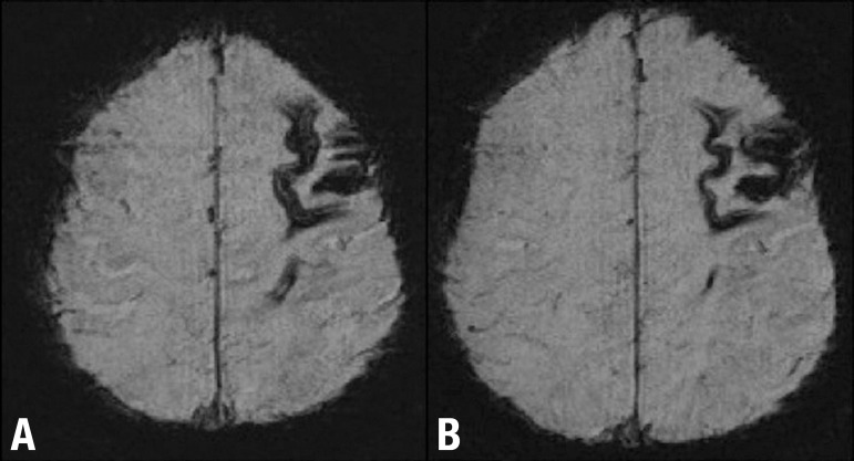
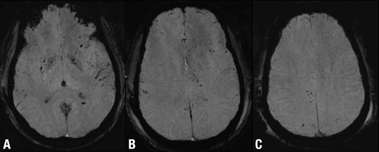
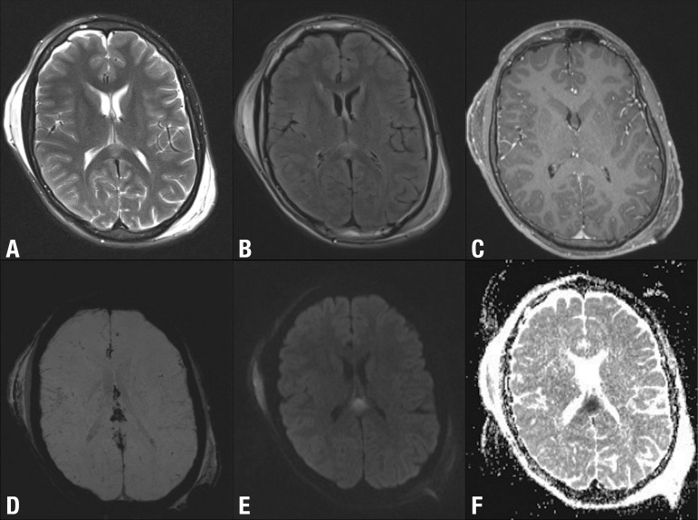
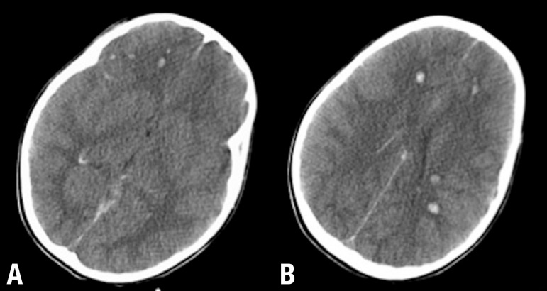
Traumatic brain injury (TBI) represents a significant public health problem in modern societies. It is primarily a consequence of traffic-related accidents and falls. Other recently recognized causes include sports injuries and indirect forces such as shock waves from battlefield explosions. TBI is an important cause of death and lifelong disability and represents the most well-established environmental risk factor for dementia. With the growing recognition that even mild head injury can lead to neurocognitive deficits, imaging of brain injury has assumed greater importance. However, there is no single imaging modality capable of characterizing TBI. Current advances, particularly in MR imaging, enable visualization and quantification of structural and functional brain changes not hitherto possible. In this review, we summarize data linking TBI with dementia, emphasizing the imaging techniques currently available in clinical practice along with some advances in medical knowledge.
O traumatismo cranioencefálico (TCE) representa um importante problema de saúde pública nas sociedades modernas. As suas principais causas são: os acidentes de trânsito e as quedas. O traumatismo leve e repetido relacionado com os esportes de contato ou o traumatismo relacionado com as ondas de choque provenientes de explosões em cenário de guerra são hoje reconhecidas como importantes causas de TCE. A mortalidade e morbilidade associada ao TCE é considerável. TCE representa o fator de risco ambiental melhor reconhecido para o desenvolvimento de demência. Com o reconhecimento recente de que até o TCE leve pode determinar déficts cognitivos, os estudos de imagem adquiriram grande importância neste contexto. Contudo, não está definido qual o melhor estudo de imagem para caracterizar o TCE. Avanços tecnológicos, como a ressonância magnética, permitem atualmente identificar e quantificar alterações intra-parenquimatosas estruturais e funcionais, não detectáveis nos estudos convencionais. Neste artigo os autores resumem os estudos que relacionam TCE e demência, dando particular ênfase às técnicas de imagem atualmente disponíveis na prática clínica, bem como alguns avanços nos métodos de imagem ainda limitados ao plano da investigação.
Keywords: chronic traumatic encephalopathy; craniocerebral trauma; dementia; magnetic resonance; post-concussion syndrome.
Conflict of interest statement
Disclosure: The authors report no conflits of interest.
Figures


References
-
- Parizel PM, Ozsarlak, Van Goethem JW, et al. Imaging findings in diffuse axonal injury after closed head trauma. Eur Radiol. 1998;8:960–965. - PubMed
-
- Stein SC, Ross SE. Clinical predictors of abnormality disclosed by computed tomography after mild head trauma. Neurosurgery. 1993;33:339–340. - PubMed
-
- Moran SG, McCarthy MC, Uddin DE, Poelstra RJ. Predictors of positive CT scans in the trauma patient with minor head injury. Am Surg. 1994;60:533–535. discussion535-536. - PubMed
-
- Duus BR, Lind B, Christensen H, Nielsen OA. The role of neuroimaging in the initial management of patients with minor head injury. Ann Emerg Med. 1994;23:1279–1283. - PubMed
Publication types
LinkOut - more resources
Full Text Sources
