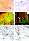
Cell junction protein armadillo repeat gene deleted in velo-cardio-facial syndrome is expressed in the skin and colocalizes with autoantibodies of patients affected by a new variant of endemic pemphigus foliaceus in Colombia
- PMID: 29214101
- PMCID: PMC5718118
- DOI: 10.5826/dpc.0704a02
Cell junction protein armadillo repeat gene deleted in velo-cardio-facial syndrome is expressed in the skin and colocalizes with autoantibodies of patients affected by a new variant of endemic pemphigus foliaceus in Colombia
Abstract
Background: We previously described a new variant of endemic pemphigus foliaceus in El Bagre, Colombia, South America (El Bagre-EPF, or pemphigus Abreu-Manu). El Bagre-EPF differs from other types of EPF clinically, epidemiologically, immunologically and in its target antigens. We reported the presence of patient autoantibodies colocalizing with armadillo repeat gene deleted in velo-cardio-facial syndrome (ARVCF), a catenin cell junction protein colocalizing with El Bagre-EPF autoantibodies in the heart and within pilosebaceous units along their neurovascular supply routes. Here we investigate the presence of ARVCF in skin and its possibility as a cutaneous El Bagre-EPF antigen.
Methods: We used a case-control study, testing sera of 45 patients and 45 controls via direct and indirect immunofluorescence (DIF/IIF), confocal microscopy, immunoelectron microscopy and immunoblotting for the presence of ARVCF and its relationship with El Bagre-EPF autoantibodies in the skin. We also immunoadsorbed samples with desmoglein 1 (Dsg1) ectodomain (El Bagre-EPF antigen) by incubating with the positive ARVCF samples from DIF and IIF.
Results: ARVCF was expressed in all the samples from the cases and controls. Immunoadsorption with Dsg1 on positive ARVCF immunofluorescence DIF/IIF cases showed that the immune response was present against non-desmoglein 1 antigen(s). Overall, 40/45 patients showed colocalization of their autoantibodies with ARVCF in the epidermis; no controls from the endemic area displayed colocalization.
Conclusions: We demonstrate that ARVCF is expressed in many areas of human skin, and colocalizes with the majority of El Bagre-EPF autoantibodies as a putative antigen.
Keywords: ARCVF; autoimmunity; cell junctions; endemic pemphigus foliaceus; skin.
Conflict of interest statement
Competing interests: The authors have no conflicts of interest to disclose.
Figures

Similar articles
-
Patients with a new variant of endemic pemphigus foliaceus have autoantibodies against arrector pili muscle, colocalizing with MYZAP, p0071, desmoplakins 1 and 2 and ARVCF.Clin Exp Dermatol. 2017 Dec;42(8):874-880. doi: 10.1111/ced.13214. Epub 2017 Oct 15. Clin Exp Dermatol. 2017. PMID: 29034528
-
Patients affected by a new variant of endemic pemphigus foliaceus have autoantibodies colocalizing with MYZAP, p0071, desmoplakins 1-2 and ARVCF, causing renal damage.Clin Exp Dermatol. 2018 Aug;43(6):692-702. doi: 10.1111/ced.13566. Epub 2018 May 16. Clin Exp Dermatol. 2018. PMID: 29768670
-
Patterns of Antinuclear Antibodies in a New Variant of Endemic Pemphigus in El Bagre, Colombia, Colocalizing with Antigens against MIZAP, ARVCF, p0071, and Desmoplakins I and II.J Appl Lab Med. 2022 Oct 29;7(6):1366-1378. doi: 10.1093/jalm/jfac050. J Appl Lab Med. 2022. PMID: 35899599
-
Rare clinical form in two patients affected by a new variant of endemic pemphigus in northern Colombia.Skinmed. 2004 Nov-Dec;3(6):317-21. doi: 10.1111/j.1540-9740.2004.03514.x. Skinmed. 2004. PMID: 15538080 Review.
-
Update on fogo selvagem, an endemic form of pemphigus foliaceus.J Dermatol. 2015 Jan;42(1):18-26. doi: 10.1111/1346-8138.12675. J Dermatol. 2015. PMID: 25558948 Free PMC article. Review.
Cited by
-
Fine mapping spatiotemporal mechanisms of genetic variants underlying cardiac traits and disease.Nat Commun. 2023 Feb 28;14(1):1132. doi: 10.1038/s41467-023-36638-2. Nat Commun. 2023. PMID: 36854752 Free PMC article.
-
The Pericardium Cells Junctions Are a Target for Autoantibodies of Patients Affected by a Variant of Endemic Pemphigus Foliaceus in El Bagre and Surrounding Municipalities in Colombia, South America.Diagnostics (Basel). 2025 Apr 10;15(8):964. doi: 10.3390/diagnostics15080964. Diagnostics (Basel). 2025. PMID: 40310349 Free PMC article.
-
Post translational Modifications and Protein-Protein Interactome of Endemic Pemphigus in El Bagre, Colombia: A New Variant Analysis.Dermatol Pract Concept. 2025 Jul 31;15(3):5097. doi: 10.5826/dpc.1503a5097. Dermatol Pract Concept. 2025. PMID: 40790431 Free PMC article.
References
-
- Diaz LA, Sampaio SA, Rivitti EA, et al. Endemic pemphigus foliaceus (fogo selvagem). I. Clinical features and immunopathology. J Am Acad Dermatol. 1989;20(4):657–659. - PubMed
-
- Ortega-Loayza AG, Ramos W, Gutierrez EL, et al. Endemic pemphigus foliaceus in the Peruvian Amazon. Clin Exp Dermatol. 2013;38(6):594–600. - PubMed
-
- Castro RM, Proença NG. Similarities and differences between Brazilian wild fire and pemphigus foliaceus Cazenave. Hautarzt. 1982;33(11):574–577. - PubMed
-
- Morini JP, Jomaa B, Gorgi Y, et al. Pemphigus foliaceus in young women. An endemic focus in the Sousse area of Tunisia. Arch Dermatol. 1993;129(1):69–73. - PubMed
-
- Abreu-Velez AM, Hashimoto T, Bollag W, et al. A unique form of endemic pemphigus in Northern Colombia. J Am Acad Dermatol. 2003;49(4):599–608. - PubMed
LinkOut - more resources
Full Text Sources
Other Literature Sources
