. 2018 Nov;25(1):1-11.
doi: 10.1080/10717544.2017.1399301.

Enhanced efficacy of curcumin with phosphatidylserine-decorated nanoparticles in the treatment of hepatic fibrosis

Affiliations

Enhanced efficacy of curcumin with phosphatidylserine-decorated nanoparticles in the treatment of hepatic fibrosis

Ji Wang et al. Drug Deliv. 2018 Nov.

Abstract

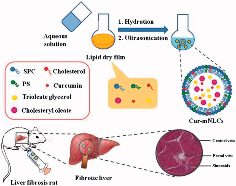
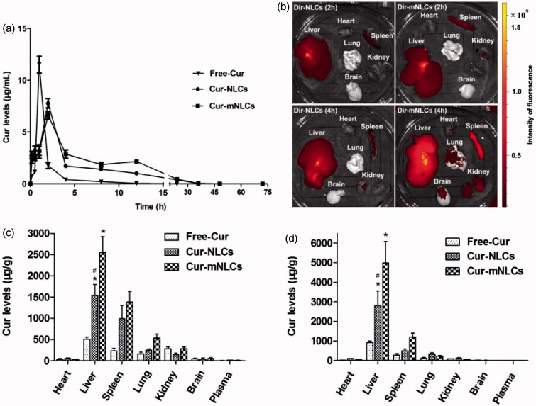
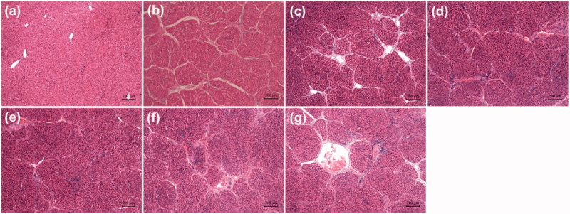
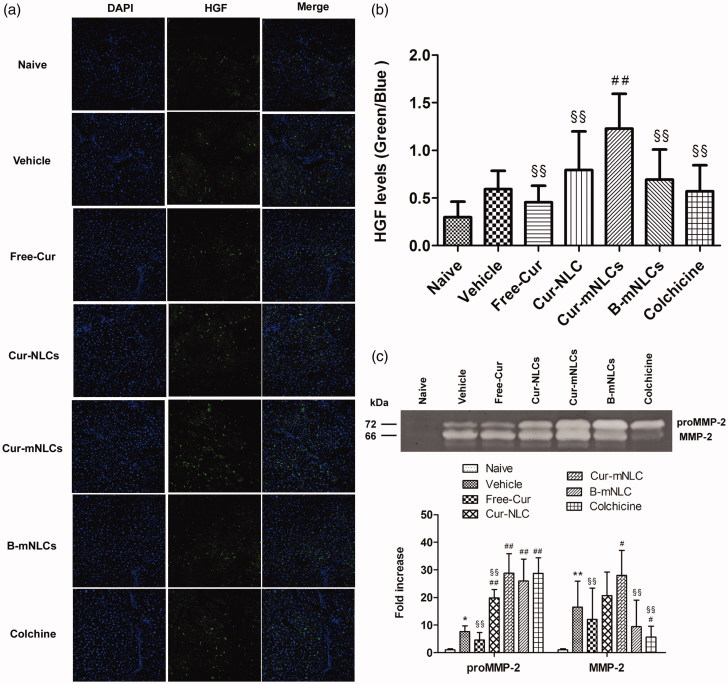
Hepatic macrophages have been considered as a therapeutic target for liver fibrosis treatment, and phosphatidylserine (PS)-containing nanoparticles are commonly used to mimic apoptotic cells that can specifically regulate macrophage functions, resulting in anti-inflammatory effects. This study was designed to test the efficacy of PS-modified nanostructured lipid carriers (mNLCs) containing curcumin (Cur) (Cur-mNLCs) in the treatment of liver fibrosis in a rat model. Carbon tetrachloride-induced liver fibrosis in rats was used as an experimental model, and the severity of the disease was examined by both biochemical and histological methods. Here, we showed that mNLCs were spherical nanoparticles with decreased negative zeta potentials due to PS decoration, and significantly increased both mean residence time and area under the curve of Cur. In the rats with liver fibrosis, PS-modification of NLCs enhanced the nanoparticles targeting to the diseased liver, which was evidenced by their highest accumulation in the liver. As compared to all the controls, Cur-mNLCs were significantly more effective at reducing the liver damage and fibrosis, which were indicated by in Cur-mNLCs-treated rats the least increase in liver enzymes and pro-inflammatory cytokines in the circulation, along with the least increase in collagen fibers and alpha smooth muscle actin and the most increased hepatocyte growth factors (HGF) and matrix metalloprotease (MMP) two in the livers. In conclusion, PS-modified NLCs nanoparticles prolonged the retention time of Cur, and enhanced its bioavailability and delivery efficiency to the livers, resulting in reduced liver fibrosis and up-regulating hepatic expression of HGF and MMP-2.

Keywords: Phosphatidylserine; curcumin; liver fibrosis; macrophages; nanostructured lipid carriers.


Conflict of interest statement

The authors declare no conflict of interest.

Figures

Figure 1.
Figure 1.
A schematic diagram of preparation procedures and liver targeting properties of Cur-mNLCs.
Figure 2.
Figure 2.
PS enhanced the retention times of Cur encapsulated in NLCs in sera as well as Cur delivery by NLCs to the liver in vivo. (a) The changes of plasma concentration of Cur after i.p. injection of Free-Cur solution, Cur-NLCs, and Cur-mNLCs in rats with liver fibrosis. Data in each point were presented as mean ± SD (n = 6). (b) Ex vivo imaging of organs in rats with liver fibrosis indicated that PS decoration improved liver targeting properties of nanocarriers. Data were presented as a typical image of major organs from one rat at each time point. (c–d) Tissue distribution of Cur after i.p. injection of Free-Cur solution, Cur-NLCs and Cur-mNLCs in rats with liver fibrosis at 2 h (c) and 4 h (d). PS decoration enhanced NLCs liver targeting properties. Data were presented as mean ± SD at each point (n = 6). *p < .01, compared with Free-Cur; # p < .01, compared with Cur-mNLCs.
Figure 3.
Figure 3.
Effects of different drug formulations on the histological changes in the liver of CCl4-treated rats as shown by H&E staining. (a) Naive group, (b) Vehicle group (positive control), (c) Free-Cur group, (d) Cur-NLCs group, (e) Cur-mNLCs group, (f) B-mNLCs group, (g) Colchicine group. Data were presented as a typical microscopic view of each group.
Figure 4.
Figure 4.
Treatment with Cur-mNLCs significantly up-regulated HGF expression and activated MMP-2 secretion in the liver. (a) Immunofluorescent staining of liver slices for antifibrotic marker HGF. (b) HGF fluorescence intensity was elevated in the livers of rats treated with different formulations in comparison with those in Vehicle group. ##p < .01, compared with Vehicle group, §§p < .01, compared with Cur-mNLCs group (mean ± SD, n = 6). (c) Gelatin zymography assay of proMMP-2 and MMP-2 activity showed that different drug formulations could accelerate liver fibrosis resolution by promoting MMP-2 secretion. *p < .05 or **p < .01, compared with Naive group, #p < .05 or ##p < .01, compared with Vehicle group, §§p < .01, compared with Cur-mNLCs group (mean ± SD, n = 6).

References

    1. Adhyatmika A, Putri KSS, Beljaars L, Melgert BN. (2015). The elusive antifibrotic macrophage. Front Med (Lausanne) 2:81. - PMC - PubMed
    1. Affò S, Rodrigo-Torres D, Blaya D, et al. (2015). Chemokine receptor Ccr6 deficiency alters hepatic inflammatory cell recruitment and promotes liver inflammation and fibrosis. Plos One 10:e0145147. - PMC - PubMed
    1. Anand P, Kunnumakkara AB, Newman RA, Aggarwal RR. (2007). Bioavailability of curcumin: problems and promises. Mol Pharm 4:807–18. - PubMed
    1. Bagalkot V, Badgeley MA, Kampfrath T, et al. (2015). Hybrid nanoparticles improve targeting to inflammatory macrophages through phagocytic signals. J Control Release 217:243–55. - PMC - PubMed
    1. Bagalkot V, Deiuliis JA, Rajagopalan S, Maiseyeu A. (2016). “Eat me” imaging and therapy. Adv Drug Deliv Rev 99:2–11. - PMC - PubMed

MeSH terms