Clinical Trial
. 2017 Dec 7;7(1):17188.
doi: 10.1038/s41598-017-17568-8.

High Dose Parenteral Ascorbate Inhibited Pancreatic Cancer Growth and Metastasis: Mechanisms and a Phase I/IIa study

Affiliations
Clinical Trial

High Dose Parenteral Ascorbate Inhibited Pancreatic Cancer Growth and Metastasis: Mechanisms and a Phase I/IIa study

Kishore Polireddy et al. Sci Rep. .

Abstract

Pancreatic cancer is among the most lethal cancers with poorly tolerated treatments. There is increasing interest in using high-dose intravenous ascorbate (IVC) in treating this disease partially because of its low toxicity. IVC bypasses bioavailability barriers of oral ingestion, provides pharmacological concentrations in tissues, and exhibits selective cytotoxic effects in cancer cells through peroxide formation. Here, we further revealed its anti-pancreatic cancer mechanisms and conducted a phase I/IIa study to investigate pharmacokinetic interaction between IVC and gemcitabine. Pharmacological ascorbate induced cell death in pancreatic cancer cells with diverse mutational backgrounds. Pharmacological ascorbate depleted cellular NAD+ preferentially in cancer cells versus normal cells, leading to depletion of ATP and robustly increased α-tubulin acetylation in cancer cells. While ATP depletion led to cell death, over-acetylated tubulin led to inhibition of motility and mitosis. Collagen was increased, and cancer cell epithelial-mesenchymal transition (EMT) was inhibited, accompanied with inhibition in metastasis. IVC was safe in patients and showed the possibility to prolong patient survival. There was no interference to gemcitabine pharmacokinetics by IVC administration. Taken together, these data revealed a multi-targeting mechanism of pharmacological ascorbate's anti-cancer action, with minimal toxicity, and provided guidance to design larger definitive trials testing efficacy of IVC in treating advanced pancreatic cancer.


Conflict of interest statement

The authors declare that they have no competing interests.

Figures

Figure 1
Figure 1
Ascorbate inhibited pancreatic cancer growth and metastasis in vitro. (A) Dose responses of pancreatic cancer cells and normal cells to ascorbate. Cells were exposed to 0–20 mM ascorbate and cell viability was detected at 24 h by MTT assays. +cat, pre-incubation with 600 U/ml catalase. Data represents Mean ± SD of 3 experiments each done in triplicates. (B) Colony formation of pancreatic cancer cells under ascorbate treatment. Cells were exposed to 5 mM of ascorbate and colonies were counted at 21–28 days. Data shows % of colonies relative to untreated control (Mean ± SD of ≥3 replicates). (C) Matrigel invasion assays for pancreatic cancer cell migration and invasion. PANC-1 cells were seeded at 1 × 104 cells/insert and were exposed to 1 mM ascorbate (Asc). Cell migration (without Matrigel) and invasion (with Matrigel) were detected at 24 hrs. Bar graph shows the average number of migrated/invaded cells per field. Data represents ≥ 3 experiments each done in triplicates. (D) qRT-PCR for changes in EMT markers. PANC-1 cells were treated with 1.5 mM Asc for 24 hrs. Data was normalized to 18s rRNA, and then compared to control PANC-1 cells for fold change. Data represent Mean ± SD of 2–6 independent experiments. (E) Western blot in PANC-1 cells showing expression of E-cadherin (E-Cad), vimentin (Vim) and Snail after ascorbate treatment. Vinculin was a loading control. Left panel shows representative blots. Bar graph shows relative band density normalized to vinculin, analyzed by Image J. (F) qRT-PCR for MMP-2 mRNA expression in PANC-1 cells treated with ascorbate for 24 hrs. Data represents Mean ± SD of 3 independent experiments. (G) Gelatin zymography assay for MMP-2 enzymatic activity after ascorbate treatment. After ascorbate treatment cell lysate were used for RNA isolation, reverse transcription and qRT-PCR. Supernatant media was used for detection of MMP-2 activity using gelatin zymography. Data represents Mean ± SD of 3 independent experiments.
Figure 2
Figure 2
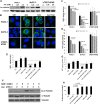
Ascorbate depleted NAD+ in pancreatic cancer cells and enhanced tubulin acetylation. (A) Western blot analysis of acetylated α-tubulin in pancreatic cancer cells (BxPC-3 and PANC-1), and an immortalized non-cancerous pancreatic ductal epithelial cell line hTERT-HPNE. Cells were treated for 4 hrs. (B) Immunofluorescence with confocal microscopy showing acetylated α-tubulin in cells treated with ascorbate (Asc) 2.5 mM or hydrogen peroxide (H2O2) 500 µM for 4 hrs. Asc + Cat, co-treatment of ascorbate and 600 U/ml catalase for 4 hrs. Cell nuclei were counter-stained blue with hoechst33342 (2 mg/mL). (C) Changes of NAD+ and (D) ATP in PANC-1, BxPC-3 and hTERT-HPNE cells treated with Asc for 4 hrs. NAD+ and ATP were detected by HPLC-UV analysis and normalized to protein contents. Data represents Mean ± SD of 2–4 independent experiments. (E) Supplementation of NAD+ protected PANC-1 cellular NAD+ levels with Asc treatment. (F) Western blot showing tubulin acetylation was prevented with supplementation of NAD+. (G) ATP in PANC-1 was protected with supplementation of NAD+. (H) Colony formation was protected with supplementation of NAD+. PANC-1 cells were pre-incubated with NAD+ for 30 min, and then treated with Asc and seeded into 2-layer soft agar for colony formation. Colonies were counted after 21 days of incubation. NAD+, tubulin acetylation, and ATP were detected at 4 h of treatment. Data represents Mean ± SD of ≥3 independent experiments.
Figure 3
Figure 3
Ascorbate over-stabilized polymerized tubulin by increased tubulin acetylation. (A) Tubulin Polymerization detected by native PAGE in PANC-1 and BxPC-3 cells. After 4 h of Asc or paclitaxel treatment, cells were lysed in RIPA buffer and subject to native PAGE. α-tubulin acetylation was confirmed by SDS PAGE and western blot as shown in the bottom panels. MW, molecular weight in kDa. (B) Cold induced microtubule depolymerization assay in PANC-1 and BxPC-3 pancreatic cancer cells. After Asc treatment for 4 h cell lysates were exposed to either 37 °C or sit on ice (4 °C) for indicated time, and were then subject to native PAGE. (C) Correlation between tubulin acetylation and cell death induced by ascorbate. All results are representatives of ≥3 independent experiments.
Figure 4
Figure 4
Ascorbate inhibited pancreatic cancer growth and metastasis in vivo. (A) Bioluminescence images of mice bearing orthotopic pancreatic xenografts treated with ascorbate (Asc), gemcitabine (Gem) or the combination of Asc+Gem. Day 0 indicated the beginning of treatment which was 2 weeks post orthotopic injection of luciferase expressing PANC-1-Leu cells into mouse pancreas. Day 45 was the end of the experiment. Asc, ascorbate treatment at intraperitoneal dose of 4 g/kg/day. Gem, gemcitabine at intraperitoneal dose of 40 mg/kg/week. Control (Ctrl) mice were treated with saline that had the same osmolarity as the ascorbate injections. (B) Total tumor burden per mouse by imaging was quantified as photons/sec/cm2 (Mean ± SEM). (C) Total tumor weight (Mean ± SEM), and (D) number of metastatic lesions in each mouse, determined by necropsy at Day 45. (E) Immunohistochemical analysis of proliferating cell nuclear antigen (PCNA) with formalin fixed tumor samples. Bar graph (right) represents the average number of PCNA positive cells per field. 15 fields from 3 different tumors from each group were analyzed. (F) Histological analysis of mitosis on H&E stained tumor slices. Bar graph (right) shows mitotic index, which was the average number of mitoses from 4 separate fields. Tumors from 4 mice in each group were examined. (G) Masson’s trichrome staining for collagen content in tumor tissues. Collagen was stained blue, and cytoplasm pink. Bar graph represents Mean ± SD of % area collagen/cross section. 15 fields from 3 different tumors from each group were analyzed. (H) H&E staining for analysis of desmoplasia in mouse tumor tissues. Bar graph shows desmoplasia represented as % of area contains desmoplastic response. Tumors from 4 mice in each group were examined (Mean ± SD). (I) Masson’s trichrome staining for collagen and fibrosis of livers from control and ascorbate treated mice. No collagen or fibrosis was seen. (J) qRT-PCR detection of 24 kinds of collagen transcripts in mouse tumor samples. Five tumors from each group were detected in duplicates. Bar shows fold changes compared to control mice in Mean ± SD. *P < 0.05; **P < 0.01; and ***P < 0.001. (K) Western blot in mouse tumor samples showing changes in CK-19 and Snail. Vinculin was used as loading control. (L) Immunohistochemistry in mouse tumor samples showing α-tubulin acetylation. Bar graph represents Mean ± SD of % area of positive staining for acetylated α-tubulin per cross section. 11–17 fields from 3 different tumors from each group were analyzed. (M) A simplified scheme showing mechanisms of ascorbate inhibiting pancreatic cancer growth and metastasis.
Figure 5
Figure 5
Overall survival (OS) and progression free survival (PFS) of the 12 participants who completed phase IIa study. Dotted lines showed median overall survival.
Figure 6
Figure 6
Pharmacokinetic parameters of gemcitabine, dFdU and ascorbate when IVC and gemcitabine were used alone or in combination. Cmax, AUC and t ½ of gemcitabine (A–C), dFdU (D–F) and ascorbate (G–I) were shown. Cmax and AUC were normalized to dose. Line graphs show parameters obtained for each subject from single drug administration (left) and combination administration (right). Box plots denote the median and the 5th, 25th, 75th, and 95th percentiles for all patients, and the outlying values.

References

    1. American Cancer Society. Cancer Facts and Figures 2017 (2017).
    1. Burris H, III, et al. Improvements in survival and clinical benefit with gemcitabine as first-line therapy for patients with advanced pancreas cancer: a randomized trial. Journal of Clinical Oncology. 1997;15:2403. doi: 10.1200/JCO.1997.15.6.2403. - DOI - PubMed
    1. Renouf D, Moore M. Evolution of systemic therapy for advanced pancreatic cancer. Expert Rev Anticancer Ther. 2010;10:529–540. doi: 10.1586/era.10.21. - DOI - PubMed
    1. Kim R. FOLFIRINOX: a new standard treatment for advanced pancreatic cancer? Lancet Oncol. 2011;12:8–9. doi: 10.1016/S1470-2045(10)70237-0. - DOI - PubMed
    1. Von Hoff DD, et al. Increased survival in pancreatic cancer with nab-paclitaxel plus gemcitabine. N Engl J Med. 2013;369:1691–1703. doi: 10.1056/NEJMoa1304369. - DOI - PMC - PubMed

Publication types

MeSH terms