Review
. 2017 Dec 7;9(12):376.
doi: 10.3390/v9120376.

Host Cell Restriction Factors that Limit Influenza A Infection

Affiliations
Review

Host Cell Restriction Factors that Limit Influenza A Infection

Fernando Villalón-Letelier et al. Viruses. .

Abstract

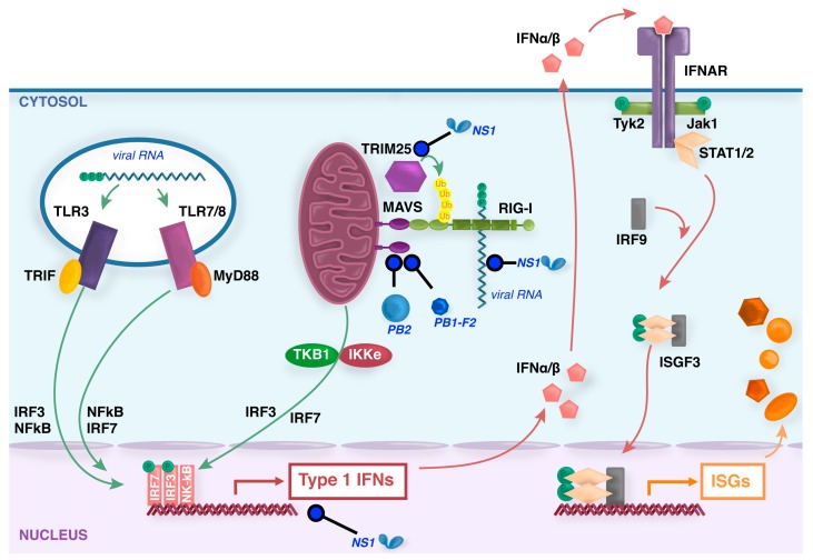
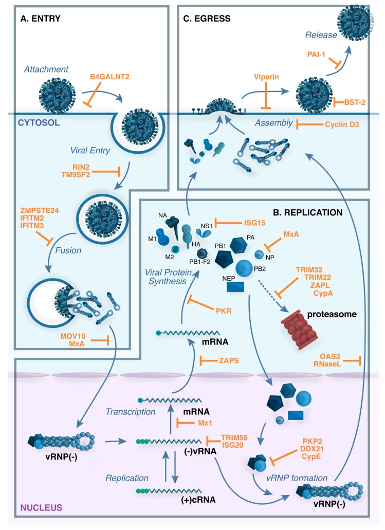
Viral infection of different cell types induces a unique spectrum of host defence genes, including interferon-stimulated genes (ISGs) and genes encoding other proteins with antiviral potential. Although hundreds of ISGs have been described, the vast majority have not been functionally characterised. Cellular proteins with putative antiviral activity (hereafter referred to as "restriction factors") can target various steps in the virus life-cycle. In the context of influenza virus infection, restriction factors have been described that target virus entry, genomic replication, translation and virus release. Genome wide analyses, in combination with ectopic overexpression and/or gene silencing studies, have accelerated the identification of restriction factors that are active against influenza and other viruses, as well as providing important insights regarding mechanisms of antiviral activity. Herein, we review current knowledge regarding restriction factors that mediate anti-influenza virus activity and consider the viral countermeasures that are known to limit their impact. Moreover, we consider the strengths and limitations of experimental approaches to study restriction factors, discrepancies between in vitro and in vivo studies, and the potential to exploit restriction factors to limit disease caused by influenza and other respiratory viruses.

Keywords: influenza; innate; interferon-stimulated gene; replication; restriction factor.


Conflict of interest statement

The authors declare no conflict of interest.

Figures

Figure 1
Figure 1
Induction of type I interferons (IFNs) by influenza viruses. During entry of influenza virus into some host cells, virions within the endosome expose viral RNA to toll-like recepotrs (TLR)3/7/8. Virus replication produces triphosphorylated vRNA and potentially dsRNA by-products that are recognized by the ubiquitously expressed cytoplasmic sensor retinoic acid inducible gene (RIG)-I. TLR signaling by the adaptors TIR-domain-containing adapter-inducing interferon-β (TRIF) or myeloid differentiation factor (MyD)88, and RIG-I signaling by the adaptor mitochondrial antiviral signaling (MAVS), trigger signal transduction cascades that result in activation of IFN regulatory factor (IRF)3/7 and NF-κB factors that translocate to the nucleus to induce synthesis of type I IFN mRNAs. Secreted type I IFNs signal through the IFN-α/β receptor complex (IFNAR) via activation of the intracellular kinases Jak1 and Tyk2, which phosphorylate the signal transducer and activator of transcription (STAT) transcription factors that, together with IRF-9, form the interferon-stimulated gene factor 3 (ISGF3) that translocates to the nucleus and activates the transcription of ISGs. In virus-infected cells, the nonstructural (NS)1 protein binds to and sequesters dsRNA to antagonize RIG-I activation, as well as interacting with various host proteins to inhibit transcription, processing or nuclear export of cellular mRNAs [20,21]. NS1 also binds to tripartite motif-containing (TRIM)25 protein and prevents essential ubiquitination of RIG-I [22]. Viral PB1-F2 [23] and PB2 proteins [24,25] inhibit MAVS function in the mitochondria.
Figure 2
Figure 2
Restriction factors block multiple stages in the influenza virus life cycle. (A) Virus attachment and entry. The influenza virus hemagglutinin (HA) attaches to host cell receptors that contain terminal α2,6-linked or α2-3-linked sialic acid moieties, and the virus enters the cell by receptor-mediated endocytosis. Cleavage of HA by cellular proteases is required to expose the HA peptide that is responsible for the fusion between the viral envelope and the endosomal membrane. Acidification of the endocytic vesicle opens the M2 ion channel, resulting in acidification of the inside of the virion, a process that is required for proper uncoating of the viral ribonucleoprotein (vRNP) complexes that contain the viral genome. Acidification of the endosome also triggers the pH-dependent fusion step that is mediated by the HA and results in the cytoplasmic release of the vRNP complexes; (B) Genomic transcription and replication. vRNPs translocate to the nucleus, where the RNA-dependent RNA polymerase transcribes and replicates the negative-sense viral RNA ((−)vRNA), giving rise to: (i) complementary positive-sense RNA ((+)cRNA), which is used as a template to generate more vRNA; and (ii) viral mRNAs, which are exported to the cytoplasm for translation; (C) Virus assembly and budding. Viral proteins that are needed in replication and transcription are translocated back to the nucleus, and progeny vRNPs are then exported to the cytoplasm for packaging. Viral HA, NA and M2 are transported by the trans-Golgi secretory pathway to the plasma membrane, where M1 assists in the formation of mature virus particles. Virions then bud from the surface of infected cells and are released by the enzymatic activity of the viral neuraminidase (NA), which cleaves sialic acid from cell-surface glycoproteins and glycolipids. Examples of cellular restriction factors with known antiviral activity against influenza viruses are shown in orange.

References

    1. Tong S., Zhu X., Li Y., Shi M., Zhang J., Bourgeois M., Yang H., Chen X., Recuenco S., Gomez J., et al. New world bats harbor diverse influenza A viruses. PLoS Pathog. 2013;9:e1003657. doi: 10.1371/journal.ppat.1003657. - DOI - PMC - PubMed
    1. Taubenberger J.K., Morens D.M. 1918 Influenza: The mother of all pandemics. Emerg. Infect. Dis. 2006;12:15–22. doi: 10.3201/eid1209.05-0979. - DOI - PMC - PubMed
    1. Schotsaert M., Garcia-Sastre A. Inactivated influenza virus vaccines: The future of TIV and QIV. Curr. Opin. Virol. 2017;23:102–106. doi: 10.1016/j.coviro.2017.04.005. - DOI - PMC - PubMed
    1. Houser K., Subbarao K. Influenza vaccines: Challenges and solutions. Cell Host Microbe. 2015;17:295–300. doi: 10.1016/j.chom.2015.02.012. - DOI - PMC - PubMed
    1. Bright R.A., Medina M.J., Xu X., Perez-Oronoz G., Wallis T.R., Davis X.M., Povinelli L., Cox N.J., Klimov A.I. Incidence of adamantane resistance among influenza A (H3N2) viruses isolated worldwide from 1994 to 2005: A cause for concern. Lancet. 2005;366:1175–1181. doi: 10.1016/S0140-6736(05)67338-2. - DOI - PubMed

Substances

LinkOut - more resources