. 2017 Dec 7;18(12):2654.
doi: 10.3390/ijms18122654.

RSV Infection in Human Macrophages Promotes CXCL10/IP-10 Expression during Bacterial Co-Infection

Affiliations

RSV Infection in Human Macrophages Promotes CXCL10/IP-10 Expression during Bacterial Co-Infection

Daniela Machado et al. Int J Mol Sci. .

Abstract

Respiratory syncytial virus (RSV), a major etiologic agent of acute lower respiratory infection constitutes the most important cause of death in young children worldwide. Viral/bacterial mixed infections are related to severity of respiratory inflammatory diseases, but the underlying mechanisms remain poorly understood. We have previously investigated the intracellular mechanisms that mediate the immune response in the context of influenza virus/Streptococcus pneumoniae (Sp) co-infection using a model of human monocyte-derived macrophages (MDMs). Here, we set up and characterized a similar model of MDMs to investigate different scenarios of RSV infection and co-infection with Sp. Our results suggest that Sp contributes to a faster and possibly higher level of CXCL10/IP-10 expression induced by RSV infection in human MDMs.

Keywords: Streptococcus pneumoniae (Sp); acute lower respiratory infection; co-infection; macrophages; respiratory syncytial virus (RSV).


Conflict of interest statement

The authors declare no conflict of interest. The founding sponsors had no role in the design of the study; in the collection, analyses, or interpretation of data; in the writing of the manuscript; or in the decision to publish the results.

Figures
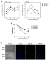
Figure 1
Figure 1
Characterization of RSV infection in human monocyte-derived macrophages (MDMs). Monocytes were isolated from human peripheral blood mononuclear cells (PBMCs) and differentiated into macrophages as previously described (1). RSV (Lon strain, ATCC VR-26) was propagated in HEp2 cells and isolated as described previously [40]. (A) THP-1 or MDMs cells were exposed to RSV Long at different multiplicity of infection (MOI, 0.1, 1 and 10) to investigate the kinetics of infection over time. Uninfected control replicates were exposed to cell culture medium. At different time points, total cell extracts were harvested and analyzed by multiplex real-time RT-qPCR to quantify RSV (F fusion gene). The RT-PCR reaction was performed using the AgPath-ID™ One-Step RT-PCR kit (Life Technologies; Carlsbad, CA, USA) according to the manufacturer’s instructions. Samples with a cycle threshold (Ct) value of ≥40 were recorded as negative. A standard curve was prepared using serially diluted RNA extracts from a known quantity and used to calculate genomic copies/mL; (B) In the same experimental condition, cell survival in infected MDMs was monitored using flow cytometry, using the FITC/Annexin V apoptosis detection kit (BD Biosciences), according to the manufacturer’s instructions. Percent survival is expressed compared to viable cells measured at T = 0 hpi; (C) Immunofluorescence microscopy of MDMs infected by RSV (MOI of 1) at 8 hpi. Magnification ×20. Immunofluorescence protocol was performed as previously published [41]. RSV F antigen (Green), Nuclei stained by DAPI (Blue). Each experiment was performed in duplicate, from two separate experiments.
Figure 2
Figure 2
Characterization of RSV/Sp mixed infection in human monocyte-derived macrophages (MDMs). Human MDMs were infected by RSV at an MOI of 1. After 4 h of viral infection, cells were mock-infected or infected by Sp at 1 CFU/cell. Encapsulated Streptococcus pneumoniae serotype 14 was obtained from the National Reference Center for Streptococci (Department of Medical Microbiology, Aachen, Germany). Pneumococci were opsonized in anti-pneumococcal immune serum and incubated for 30 min at 37 °C prior to infections for all experiments. MDM cells showed significant loss of viability following overnight incubation with S. pneumoniae, and gentamicin (10 µg/mL) was therefore added to all wells at the 2-h time point to prevent bacterial overgrowth and loss of cell viability. (A) At different time points, total cell extracts were harvested and analyzed by multiplex real-time RT-qPCR to quantify RSV; (B) RSV viral production in supernatants was performed by quantification of viral titers (TCID 50/mL) with a limit-dilution assay and using the Reed & Muench statistical method. Results are expressed as log10 TCID50/mL values; (C) In the same experimental condition, cell survival in infected MDMs was monitored using flow cytometry, with the FITC/Annexin V apoptosis detection kit (BD Biosciences), according to the manufacturer’s instructions. Percent survival is expressed compared to viable cells measured at T = 0 hpi; (D,E) Co-infection alters the timing and extent of IP-10 expression in human monocyte-derived macrophages (MDMs). Human MDMs were infected by RSV at an MOI of 1. After 4 h of viral infection, cells were mock-infected or infected by Sp at 1 CFU/cell. At different time points, total cell extracts were harvested and analyzed by multiplex real-time RT-qPCR to quantify CXCL10 mRNA levels. Total mRNA was purified from transfected and infected MDMs using the RNeasy kit (Qiagen) and specific primers were used to amplify CXCL10 (F: 5′-GTGGCATTCAAGGAGTACCTC-3′, R: 5′-TGATGGCCTTCGATTCTGGATT-3′) and β-Actin for normalization (F: 5′-CTCTTCCAGCCTTCCTTCCT-3′, R: 5′-AGCACTGTGTTGGCGTACAG-3′); (E) Similar strategy with different combinations of RSV and Sp, at 8 hpi. Each experiment was performed in duplicate, from two separate experiments.

Similar articles

Cited by

References

    1. Stupka J.E., Mortensen E.M., Anzueto A., Restrepo M.I. Community-acquired pneumonia in elderly patients. Aging Health. 2009;5:763–774. doi: 10.2217/ahe.09.74. - DOI - PMC - PubMed
    1. Wong J.L., Evans S.E. Bacterial Pneumonia in Patients with Cancer: Novel Risk Factors and Management. Clin. Chest Med. 2017;38:263–277. doi: 10.1016/j.ccm.2016.12.005. - DOI - PMC - PubMed
    1. Vakil E., Evans S.E. Viral Pneumonia in Patients with Hematologic Malignancy or Hematopoietic Stem Cell Transplantation. Clin. Chest Med. 2017;38:97–111. doi: 10.1016/j.ccm.2016.11.002. - DOI - PMC - PubMed
    1. Johansson N., Kalin M., Tiveljung-Lindell A., Giske C.G., Hedlund J. Etiology of community-acquired pneumonia: Increased microbiological yield with new diagnostic methods. Clin. Infect. Dis. 2010;50:202–209. doi: 10.1086/648678. - DOI - PMC - PubMed
    1. Honkinen M., Lahti E., Osterback R., Ruuskanen O., Waris M. Viruses and bacteria in sputum samples of children with community-acquired pneumonia. Clin. Microbiol. Infect. 2012;18:300–307. doi: 10.1111/j.1469-0691.2011.03603.x. - DOI - PMC - PubMed

Substances

LinkOut - more resources