. 2017 Dec 6;96(5):1024-1032.e3.
doi: 10.1016/j.neuron.2017.11.013.

ApoE4 Accelerates Early Seeding of Amyloid Pathology

Affiliations

ApoE4 Accelerates Early Seeding of Amyloid Pathology

Chia-Chen Liu et al. Neuron. .

Abstract

Accumulation and aggregation of amyloid-β (Aβ) in the brain is an initiating step in the pathogenesis of Alzheimer's disease (AD). The ε4 allele of apolipoprotein E (apoE) gene is the strongest genetic risk factor for late-onset AD. Although there is strong evidence showing that apoE4 enhances amyloid pathology, it is not clear what the critical stage(s) is during amyloid development in which apoE4 has the strongest impact. Using apoE inducible mouse models, we show that increased expression of astrocytic apoE4, but not apoE3, during the seeding stage of amyloid development enhanced amyloid deposition and neuritic dystrophy in amyloid model mice. ApoE4, but not apoE3, significantly increased brain Aβ half-life measured by in vivo microdialysis. Furthermore, apoE4 expression increased whereas apoE3 reduced amyloid-related gliosis in the mouse brains. Together, our results demonstrate that apoE4 has the greatest impact on amyloid during the seeding stage, likely by perturbing Aβ clearance and enhancing Aβ aggregation.

Keywords: Alzheimer’s disease; amyloid pathology; apolipoprotein E; seeding.


Figures

Figure 1
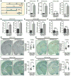
Figure 1. Characterization of Cell Type-specific and Inducible ApoE Mice. See also Figure S1
(A) The structure of the ROSA-targeting vector with the Tet-off regulation cassette for APOE and eGFP expression. The resultant mice were named iE3 and iE4. Breeding iE3 or iE4 mice with GFAP-Cre mice which removed the loxP-flanked Neor gene led to expression of apoE3 or apoE4 in astrocytes. (B, C) ApoE levels in the cortex of iE3 and iE4 mice at 3 months of age with or without GFAP-Cre were analyzed by Western blotting (B) and ELISA (C). (D) Immunohistochemical analysis of GFP for the cortical region of iE3 mice in the presence or absence of Cre. (E) The iE3 mice at 3 months of age were fed with regular (−Dox) or Dox-containing chow for 2 weeks. ApoE in the cortex was analyzed by ELISA. Data represent mean ± SEM. **, p<0.01; N.S., not significant. (F) Comparison of apoE levels in the cortex of apoE inducible amyloid model mice with apoE-TR mice at 3–4 months of age examined by ELISA. Data represent mean ± SEM. **, p<0.01. (G) Cortex and hippocampus (HPC) from apoE3 inducible (Cre+) amyloid model mice were co-immunostained for GFP which represents apoE distribution (green) and astrocyte-specific (anti-GFAP; red) or neuron-specific (anti-NeuN; red) markers. Scale bar, 100 μm.
Figure 2
Figure 2. Astrocytic ApoE4 Increases Insoluble Aβ and Amyloid Pathology When Expressed Early. See also Figure S1-S3
(A) Illustration of apoE induction paradigms at different stages of amyloid pathology. (B, C) Insoluble apoE levels in the cortex of APP/PS1 mice expressing (B) apoE3 (APP/iE3; n=17–18/group) throughout 9 months (0–9 m On) or (C) apoE4 (APP/iE4; n=13–16/group) throughout 9 months, during 0–6 months (0–6 m On; n=6–8/group) or during 6–9 months (6–9 m On; n=8–9/group) were examined by ELISA. Data represent mean ± SEM. *, p<0.05; **, p<0.01. (D–F) Insoluble Aβ40 and Aβ42 levels in the cortex of APP/PS1 mice expressing apoE4 (APP/iE4; n=13–16/group) throughout the entire 9 months, during 0–6 months (n=6–8/group) or during 6–9 months (n=8–9/group) were examined by specific Aβ ELISA. Data represent mean ± SEM. *, p<0.05; **, p<0.01; N.S., not significant. (G–J) Brain sections from 9-month-old APP/PS1 mice expressing apoE3 (APP/iE3; n=12–14/group), or apoE4 (APP/iE4) throughout the entire 9 months (n=11/group), during 0–6 months (n=9/group) or 6–9 months (n=7–8/group) were immunostained with a pan-Aβ antibody. Representative images of Aβ staining in the cortical and hippocampal regions are shown. Scale bar, 1 mm. Open circles are females; closed circles are males. Data represent mean ± SEM. *, p<0.05; **, p<0.01; N.S., not significant.
Figure 3
Figure 3. Induced ApoE4 Expression in Astrocytes Impairs ISF Aβ Clearance in the Hippocampus of APP/PS1 Mice
The ISF Aβ40 levels in APP/PS1 mice expressing apoE3 (A-B, APP/iE3; n=8/group) or apoE4 (C–D, APP/iE4; n=4/group) at the age of 3–4 months were analyzed. To assess Aβ40 half-life, the mice were treated with a γ-secretase inhibitor, and the hippocampal ISF Aβ40 levels were monitored. The slope from the individual linear regressions from log (% ISF Aβ40) versus time for each mouse was used to calculate the mean half-life (t1/2) of elimination for Aβ from the ISF (B, D). Data represent mean ± SEM. *, p<0.05; N.S., not significant.
Figure 4
Figure 4. ApoE Isoform-specific Effects on Aβ-associated Gliosis. See also Figure S4
(A–D) Brain sections from 9-month-old APP/PS1 mice expressing apoE3 (APP/iE3) or apoE4 (APP/iE4) in the astrocytes throughout the entire 9 months were immunostained with GFAP antibody (A, B) or Iba1 antibody (C, D). Scale bar, 100 μm. The immunoreactivities of GFAP or Iba1 in cortex (n=11–13/group) and hippocampus (n=9–12/group) were quantified. Data represent mean ± SEM. *, p<0.05; **, p<0.01. (E, F) The levels of GFAP, presynaptic marker synaptophysin (Syp) and postsynaptic marker PSD-95 in the cortex (n=16–18/group) of APP/PS1 mice expressing apoE3 (APP/iE3) or apoE4 (APP/iE4) in the astrocytes throughout the entire 9 months were examined by Western blotting. Data represent mean ± SEM. *, p<0.05; **, p<0.01; N.S., not significant.

Comment in

References

    1. Bajenaru ML, Zhu Y, Hedrick NM, Donahoe J, Parada LF, Gutmann DH. Astrocyte-specific inactivation of the neurofibromatosis 1 gene (NF1) is insufficient for astrocytoma formation. Mol Cell Biol. 2002;22:5100–5113. - PMC - PubMed
    1. Bales KR, Liu F, Wu S, Lin S, Koger D, DeLong C, Hansen JC, Sullivan PM, Paul SM. Human APOE isoform-dependent effects on brain beta-amyloid levels in PDAPP transgenic mice. J Neurosci. 2009;29:6771–6779. - PMC - PubMed
    1. Bales KR, Verina T, Dodel RC, Du Y, Altstiel L, Bender M, Hyslop P, Johnstone EM, Little SP, Cummins DJ, et al. Lack of apolipoprotein E dramatically reduces amyloid beta-peptide deposition. Nat Genet. 1997;17:263–264. - PubMed
    1. Bateman RJ, Munsell LY, Morris JC, Swarm R, Yarasheski KE, Holtzman DM. Human amyloid-beta synthesis and clearance rates as measured in cerebrospinal fluid in vivo. Nat Med. 2006;12:856–861. - PMC - PubMed
    1. Bateman RJ, Xiong C, Benzinger TL, Fagan AM, Goate A, Fox NC, Marcus DS, Cairns NJ, Xie X, Blazey TM, et al. Clinical and biomarker changes in dominantly inherited Alzheimer’s disease. N Engl J Med. 2012;367:795–804. - PMC - PubMed