. 2017 Dec 8;14(1):51.
doi: 10.1186/s12989-017-0232-2.

Pro-inflammatory adjuvant properties of pigment-grade titanium dioxide particles are augmented by a genotype that potentiates interleukin 1β processing

Affiliations

Pro-inflammatory adjuvant properties of pigment-grade titanium dioxide particles are augmented by a genotype that potentiates interleukin 1β processing

Sebastian Riedle et al. Part Fibre Toxicol. .

Abstract

Background: Pigment-grade titanium dioxide (TiO2) particles are an additive to some foods (E171 on ingredients lists), toothpastes, and pharma-/nutraceuticals and are absorbed, to some extent, in the human intestinal tract. TiO2 can act as a modest adjuvant in the secretion of the pro-inflammatory cytokine interleukin 1β (IL-1β) when triggered by common intestinal bacterial fragments, such as lipopolysaccharide (LPS) and/or peptidoglycan. Given the variance in human genotypes, which includes variance in genes related to IL-1β secretion, we investigated whether TiO2 particles might, in fact, be more potent pro-inflammatory adjuvants in cells that are genetically susceptible to IL-1β-related inflammation.

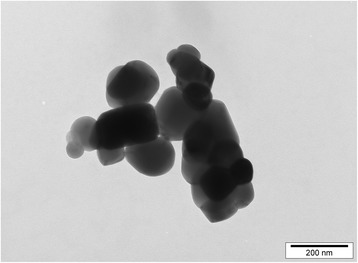
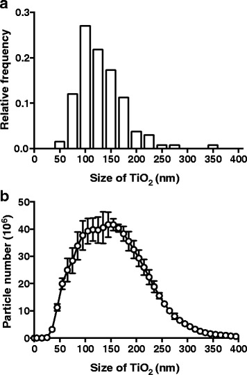
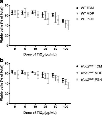
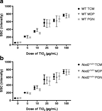
Methods: We studied bone marrow-derived macrophages from mice with a mutation in the nucleotide-binding oligomerisation domain-containing 2 gene (Nod2 m/m), which exhibit heightened secretion of IL-1β in response to the peptidoglycan fragment muramyl dipeptide (MDP). To ensure relevance to human exposure, TiO2 was food-grade anatase (119 ± 45 nm mean diameter ± standard deviation). We used a short 'pulse and chase' format: pulsing with LPS and chasing with TiO2 +/- MDP or peptidoglycan.

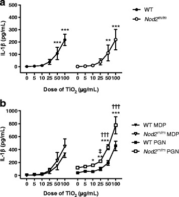
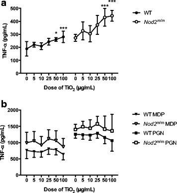
Results: IL-1β secretion was not stimulated in LPS-pulsed bone marrow-derived macrophages, or by chasing with MDP, and only very modestly so by chasing with peptidoglycan. In all cases, however, IL-1β secretion was augmented by chasing with TiO2 in a dose-dependent fashion (5-100 μg/mL). When co-administered with MDP or peptidoglycan, IL-1β secretion was further enhanced for the Nod2 m/m genotype. Tumour necrosis factor α was triggered by LPS priming, and more so for the Nod2 m/m genotype. This was enhanced by chasing with TiO2, MDP, or peptidoglycan, but there was no additive effect between the bacterial fragments and TiO2.

Conclusion: Here, the doses of TiO2 that augmented bacterial fragment-induced IL-1β secretion were relatively high. In vivo, however, selected intestinal cells appear to be loaded with TiO2, so such high concentrations may be 'exposure-relevant' for localised regions of the intestine where both TiO2 and bacterial fragment uptake occurs. Moreover, this effect is enhanced in cells from Nod2 m/m mice indicating that genotype can dictate inflammatory signalling in response to (nano)particle exposure. In vivo studies are now merited.

Keywords: E171; IL-1β; Muramyl dipeptide; NOD2; Nano; Particle; Peptidoglycan; TNF-α; TiO2.


Conflict of interest statement

Ethics approval

Collection of bone marrow from mice for this research was approved by the Grasslands Ethics Committee (Palmerston North, New Zealand), AgResearch Animal Ethics Committee, applications AE Tissue Collection 54 and 68 in compliance with the New Zealand Animal Welfare Act 1999.

Use of human blood for this research was approved by the ethics committee of the University of Cambridge (Cambridge, UK), Human Biology Research Ethics Committee, application HBREC.2015.10.

Consent for publication

Not applicable.

Competing interests

The authors declare that they have no competing interests.

Publisher’s Note

Springer Nature remains neutral with regard to jurisdictional claims in published maps and institutional affiliations.

Figures

Fig. 1
Fig. 1
Transmission electron microscopy image of TiO2 particles. Food- and pharmaceutical-grade anatase TiO2 particles were suspended in distilled water with 0.5% BSA at a concentration of 1 mg/mL. The particle suspension was analysed with transmission electron microscopy at 80 kV. A representative image is shown; scale bar = 200 nm
Fig. 2
Fig. 2
Size determination of TiO2 particles. a Food- and pharmaceutical-grade anatase TiO2 particles were suspended in distilled water with 0.5% BSA at a concentration of 1 mg/mL. The particle suspension was analysed with transmission electron microscopy at 80 kV. TiO2 particle diameters were measured with image analysis software. The distribution of the particle diameters, grouped in sizes of 25 nm, is shown as a relative frequency histogram, n = 133. b Food- and pharmaceutical-grade anatase TiO2 particles were suspended in RPMI 1640 medium with 10% FBS and 1% penicillin-streptomycin at a concentration of 1 mg/mL. TiO2 particle sizes were determined with nanoparticle tracking analysis, and the size distribution of the particles is plotted as a line graph. Data represent mean ± SD from three independent experiments
Fig. 3
Fig. 3
Viability of LPS-primed BMDMs after chasing with TiO2 +/− peptidoglycan or MDP. BMDMs from WT (a) and Nod2 m/m (b) mice were pre-stimulated for 3 h with LPS (10 ng/mL). Then BMDMs were incubated for 3 h with the indicated concentrations of TiO2 particles suspended in TCM alone (TCM), or TCM + 10 μg/mL MDP (MDP), or TCM + 10 μg/mL peptidoglycan (PGN). Cells were stained with PI and F4/80 antibody for murine macrophages and analysed with flow cytometry, and viability was determined with PI exclusion. Percentages of PI cells in relation to the total number of detected events are shown. Data represent mean ± SD from two independent experiments with three replicates each, n = 6
Fig. 4
Fig. 4
Particle uptake by LPS-primed BMDMs after chasing with TiO2 +/− peptidoglycan or MDP. BMDMs from WT (a) and Nod2 m/m (b) mice were pre-stimulated for 3 h with LPS (10 ng/mL). Then BMDMs were incubated for 3 h with the indicated concentrations of TiO2 particles suspended in TCM alone (TCM), TCM + 10 μg/mL MDP (MDP), or TCM + 10 μg/mL peptidoglycan (PGN). Cells were stained with PI and F4/80 antibody for murine macrophages and analysed with flow cytometry, and median SSC intensities of PIF4/80+ cells were recorded. Data represent mean ± SD from two independent experiments with three replicates each, n = 6
Fig. 5
Fig. 5
Correlation of SSC intensity and dark spots in bright-field images by flow and imaging cytometry. PBMCs from human blood were incubated for 24 h with 0 μg/mL, 5 μg/mL, or 10 μg/mL TiO2 particles in TCM and stained with either CD11c or CD14 antibodies for human monocytes/myeloid cells and analysed with conventional flow or imaging cytometry, respectively. a Correlation between increases in SSC MFI of CD11c+ myeloid cells identified using conventional flow cytometry and the percentages of CD14+ cells bearing dark spots in bright-field measured by imaging cytometry; Pearson correlation p < 0.01, r = 0.8424. b Representative images of cells designated ‘dark spot negative’ or ‘dark spot positive’ by imaging cytometry; scale bar = 10 μm
Fig. 6
Fig. 6
IL-1β secretion by LPS-primed BMDMs after chasing with TiO2 +/− peptidoglycan or MDP. BMDMs from WT and Nod2 m/m mice were pre-stimulated for 3 h with LPS (10 ng/mL). Then BMDMs were incubated for 3 h with the indicated concentrations of TiO2 particles suspended in TCM alone (a) or suspended in TCM + 10 μg/mL MDP (MDP) or TCM + 10 μg/mL peptidoglycan (PGN) (b). Supernatant concentrations of IL-1β were analysed by ELISA. Data represent mean ± SD from two independent experiments with three replicates each, n = 6. a Results were analysed with one-way ANOVA and Tukey’s post-hoc test; **p < 0.01, ***p < 0.001 compared to respective WT or Nod2 m/m cells incubated without TiO2. b Results were analysed with two-way ANOVA and Tukey’s post-hoc test; *p < 0.05, **p < 0.01, ***p < 0.001 for Nod2 m/m cells compared to WT cells cultured with the same TiO2 concentration, ††† p < 0.001 for WT and Nod2 m/m cells compared to respective WT or Nod2 m/m cells incubated without TiO2, p < 0.05 for Nod2 m/m cells compared to Nod2 m/m cells incubated without TiO2
Fig. 7
Fig. 7
TNF-α secretion by LPS-primed BMDMs after chasing with TiO2 +/− peptidoglycan or MDP. BMDMs from WT and Nod2 m/m mice were pre-stimulated for 3 h with LPS (10 ng/mL). Then BMDMs were incubated for 3 h with the indicated concentrations of TiO2 particles suspended in TCM alone (a) or suspended in TCM + 10 μg/mL MDP (MDP) or TCM + 10 μg/mL peptidoglycan (PGN) (b). Supernatant concentrations of TNF-α were analysed by ELISA. Data represent mean ± SD from two independent experiments with three replicates each, n = 6. a Results were analysed with one-way ANOVA and Tukey’s post-hoc test; *p < 0.05, ***p < 0.001 compared to respective WT or Nod2 m/m cells incubated without TiO2
Fig. 8
Fig. 8
IL-1β secretion by PBMCs following exposure to MSU crystals or SNPs. Secretion of IL-1β by PBMCs with (main figure) or without (inset) initial exposure to 10 ng/mL LPS for 3 h followed by exposure to MSU crystals (100 μg/mL) or SNPs (100 μg/mL) for a further 3 h. The supernatant was either collected immediately for analysis (3 h; open bars) or following a further 21 h of cell incubation in fresh (i.e. without added particles or MAMPs) TCM (3 + 21 h; black bars). All data are expressed as mean ± SEM (n = 4). Results were analysed by paired T test in comparison to baseline (B), i.e. non-particle-exposed cells; *p < 0.05 and **p < 0.01 versus respective baseline

References

    1. Shi H, Magaye R, Castranova V, Zhao J. Titanium dioxide nanoparticles: a review of current toxicological data. Part Fibre Toxicol. 2013;10:15. doi: 10.1186/1743-8977-10-15. - DOI - PMC - PubMed
    1. Kreyling WG, Holzwarth U, Schleh C, Kozempel J, Wenk A, Haberl N, et al. Quantitative biokinetics of titanium dioxide nanoparticles after oral application in rats: part 2. Nanotoxicology. 2017;11:443–453. doi: 10.1080/17435390.2017.1306893. - DOI - PubMed
    1. Lomer MCE, Hutchinson C, Volkert S, Greenfield SM, Catterall A, Thompson RPH, et al. Dietary sources of inorganic microparticles and their intake in healthy subjects and patients with Crohn's disease. Brit J Nutr. 2004;92:947–955. doi: 10.1079/BJN20041276. - DOI - PubMed
    1. Powell JJ, Faria N, Thomas-McKay E, Pele LC. Origin and fate of dietary nanoparticles and microparticles in the gastrointestinal tract. J Autoimmun. 2010;34:J226–JJ33. doi: 10.1016/j.jaut.2009.11.006. - DOI - PubMed
    1. Rompelberg C, Heringa MB, van Donkersgoed G, Drijvers J, Roos A, Westenbrink S, et al. Oral intake of added titanium dioxide and its nanofraction from food products, food supplements and toothpaste by the Dutch population. Nanotoxicology. 2016;10:1404–1414. doi: 10.1080/17435390.2016.1222457. - DOI - PubMed

MeSH terms

LinkOut - more resources