

Review
doi: 10.1016/j.otc.2017.09.002.
New Frontiers in Our Understanding of Lymphatic Malformations of the Head and Neck: Natural History and Basic Research
Affiliations
- PMID: 29217059
- PMCID: PMC5726598
- DOI: 10.1016/j.otc.2017.09.002
Item in Clipboard
Review
New Frontiers in Our Understanding of Lymphatic Malformations of the Head and Neck: Natural History and Basic Research
Otolaryngol Clin North Am.
2018 Feb.
Abstract
The future of head and neck lymphatic malformation (HNLM) evaluation and treatment is changing because of 2 decades of clinical research and recent basic science investigation. Basic science investigation using cellular biology and molecular genetics has revealed the genetic cause of some HNLMs, which has created the possibility of medical treatment specific to HNLM. This article summarizes the clinical and basic science research that will likely influence the future of HNLM assessment and treatment.
Keywords: Basic research; Head and neck; Lymphatic malformations; Natural history.
Copyright © 2017 Elsevier Inc. All rights reserved.
Figures
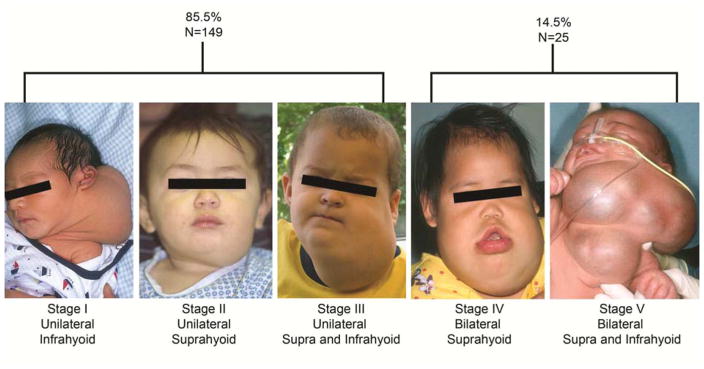
The deSerres head and neck lymphatic malformation staging system used to improve treatment outcome measurement and allow for quantitative data analysis. In a series of 174 head and neck lymphatic malformations 85.5% were stages 1–3, 14.5% were stage 4 or 5 and that in lower stage lesions surgery and sclerotherapy had the same efficacy. From: Balakrishnan K, Menezes MD, Chen BS, Magit AE, Perkins JA, et al. Primary surgery vs primary sclerotherapy for head and neck lymphatic malformations. JAMA Otolaryngol Head Neck Surg. 2014;140(1):41–45; with permission, and de Serres LM, Sie KC, Richardson MA, et al. Lymphatic malformations of the head and neck. A proposal for staging. Arch Otolaryngol Head Neck. 1995;121(5):577–582; with permission.
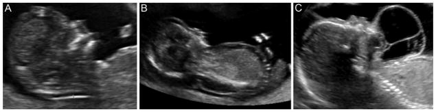
In-utero ultrasound images demonstrating (A) nuchal thickening, (B) dorsal lymphatic malformation, and (C) ventral lymphatic malformation.
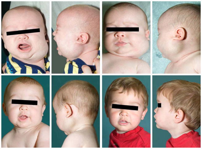
Stage 1 HNLM demonstrating regression without therapy. From left to right top row, age 2.5 months and 3 months. From left to right bottom row, age 6 months and 17 months.
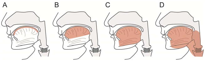
Tongue lymphatic malformation staging used to describe treatment outcomes and strategies in malformations involving the tongue (shaded area is involved with lymphatic malformation). Malformations ranged from superficial to transmural. The more extensive the malformation the poorer the treatment outcome and malformation persistence. From: Wiegand S, Eivazi B, Zimmermann AP, et al. Microcystic lymphatic malformations of the tongue: Diagnosis, classification, and treatment. Arch Otolaryngol Head Neck Surg. 2009;135(10):976–983; with permission.
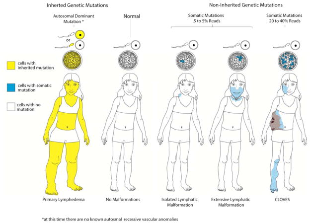
Schematic diagram of current theory of molecular genetics applied to germline and postzygotic somatic gene mutations creation of phenotype. On the left a person without malformations has a normal genome in all cells. In autosomal dominant germline inheritance all cells in the body have a mutation, shown as yellow. Somatic mutations occur after conception (i.e. zygote formation) and affect a variable number of cells in the blastomere, shown as blue. Cells with somatic mutations, by unknown mechanisms, affect one portion of the body as seen in blue. When 5–10% of cells, assuming “reads” are a surrogate measure for affected cells, are affected the involved area is small. The involved area becomes larger and more dysfunctional when more cells have that mutation.
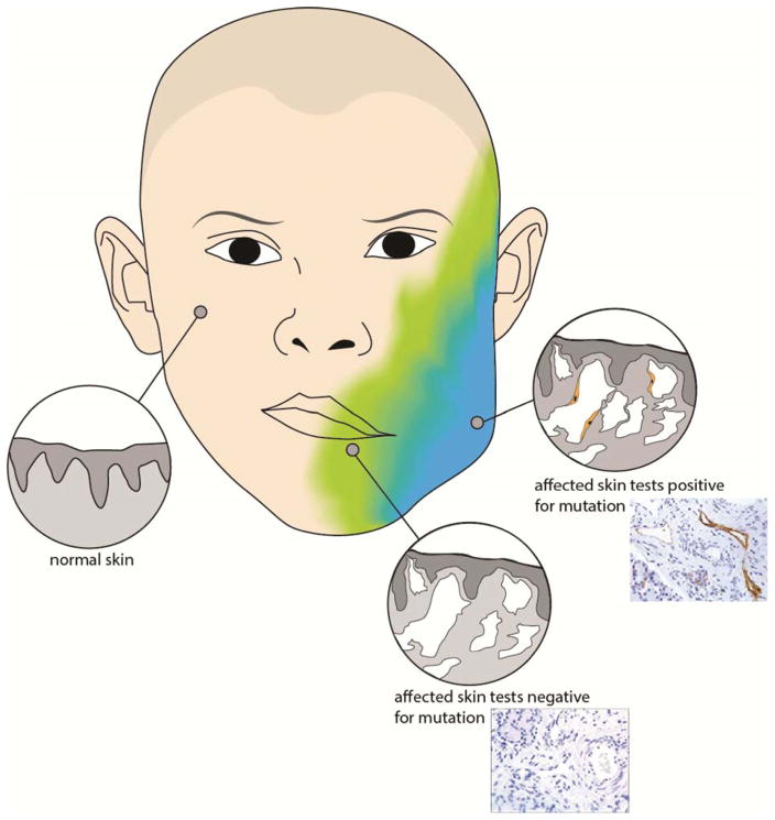
Adjacent cells interact, through mobile genomic sequences (i.e. transposable elements) and programmed cell death (i.e. apoptosis), creating an environment in which cells with somatic mutations cause neighboring genetically normal cells to exhibit mutant histologic phenotype. This may explain the occurrence and persistence of large areas of histologically abnormal lymphatic malformation tissue, schematically depicted and mirrored with HNLM tissue sections (top image with D2-40 immunostained lymphatic endothelium (brown)), while not all cells in the region have detectable mutations.
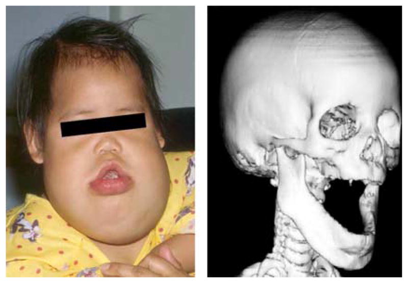
The gain-of-function post-zygotic somatic mutation in PIK3CA causes the persistent soft tissue and boney tissue overgrowth in this LM patient. Interestingly the one of the other known functions of the PIK3CA gene pathway is t-cell or lymphocyte differentiation by the mtor enzyme. This patient also has persistent lymphocytopenia which is probably related to disordered PIK3CA function.
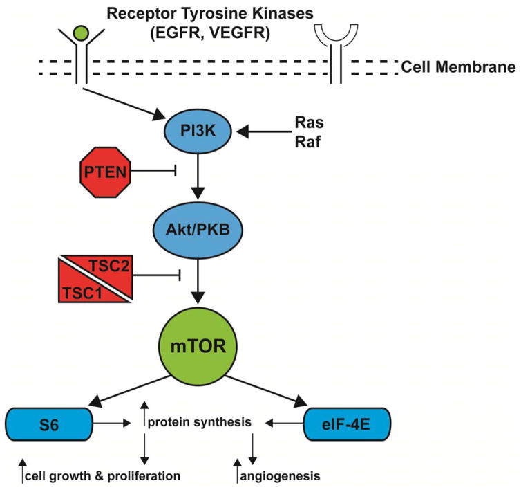
Schematic representation of PIK3CA cellular signaling pathway. Note, one of the principle functions of the mTOR enzyme is T-cell differentiation or programming.
References
-
- Luks VL, Kamitaki N, Vivero MP, et al. Lymphatic and other vascular malformative/overgrowth disorders are caused by somatic mutations in PIK3CA. [Accessed 20150330];J Pediatr. 2015 166(4):1048–54. e1–5. http://dx.doi.org/10.1016/j.jpeds.2014.12.069. - DOI - PMC - PubMed
-
- Osborn AJ, Dickie P, Neilson DE, et al. Activating PIK3CA alleles and lymphangiogenic phenotype of lymphatic endothelial cells isolated from lymphatic malformations. Hum Mol Genet. 2015;24(4):926–938. - PubMed
-
- Longstreet B, Bhama PK, Inglis AF, Jr, Saltzman B, Perkins JA. Improved airway visualization during direct laryngoscopy using self-retaining laryngeal retractors: A quantitative study. Otolaryngol Head Neck Surg. 2011;145(2):270–275. - PubMed
-
- Dighe MK, Peterson SE, Dubinsky TJ, Perkins J, Cheng E. EXIT procedure: Technique and indications with prenatal imaging parameters for assessment of airway patency. Radiographics. 2011;31(2):511–526. - PubMed
-
- Perkins JA, Maniglia C, Magit A, Sidhu M, Manning SC, Chen EY. Clinical and radiographic findings in children with spontaneous lymphatic malformation regression. Otolaryngol Head Neck Surg. 2008;138(6):772–777. - PubMed
Publication types
MeSH terms
Supplementary concepts
Grants and funding
LinkOut - more resources
Full Text Sources
Other Literature Sources
Medical
