. 2018 Jan 3:66:127-136.
doi: 10.1016/j.jbiomech.2017.11.011. Epub 2017 Nov 16.

Microstructural changes associated with osteoporosis negatively affect loading-induced fluid flow around osteocytes in cortical bone

Affiliations

Microstructural changes associated with osteoporosis negatively affect loading-induced fluid flow around osteocytes in cortical bone

Vittorio Gatti et al. J Biomech. .

Abstract

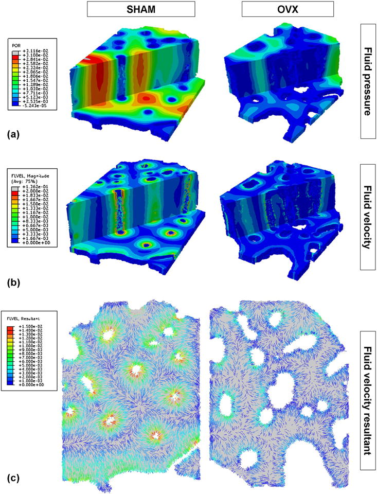
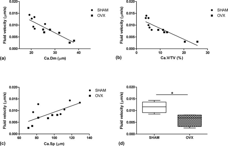
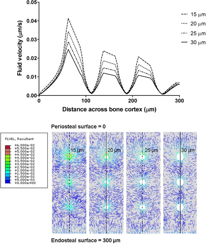
Loading-induced interstitial fluid flow in the microporosities of bone is critical for osteocyte mechanotransduction and for the maintenance of tissue health, enhancing convective transport in the lacunar-canalicular system. In recent studies, our group has reported alterations of bone's vascular porosity and lacunar-canalicular system microarchitecture in a rat model of postmenopausal osteoporosis. In this work, poroelastic finite element analysis was used to investigate whether these microstructural changes can affect interstitial fluid flow around osteocytes. Animal-specific finite element models were developed combining micro-CT reconstructions of bone microstructure and measures of the poroelastic material properties. These models were used to quantify and compare loading-induced fluid flow in the lacunar-canalicular system of ovariectomized and sham-operated rats. A parametric analysis was also used to quantify the influence of the lacunar-canalicular permeability and vascular porosity on the fluid velocity magnitude. Results show that mechanically-induced interstitial fluid velocity can be significantly reduced in the lacunar-canalicular system of ovariectomized rats. Interestingly, the vascular porosity is shown to have a major influence on interstitial fluid flow, while the lacunar-canalicular permeability influence is limited when larger than 10-20m2. Altogether our results suggest that microstructural changes associated with the osteoporotic condition can negatively affect interstitial fluid flow around osteocytes in the lacunar-canalicular system of cortical bone. This fluid flow reduction could impair mechanosensation of the osteocytic network, possibly playing a role in the initiation and progression of age-related bone loss and postmenopausal osteoporosis.

Keywords: Bone fluid flow; Lacunar-canalicular system; Osteocyte; Osteoporosis; Poroelasticity.


Conflict of interest statement

Conflict of interest statement

The authors have nothing to disclose.

Figures

Figure 1
Figure 1
Micro-CT images of (a) a whole rat tibia and (b) a proximal metaphysis cross-section from a SHAM sample, demonstrating the volume of interest selected ~1 mm distal to the growth plate in the anterior region of the tibial cortex, including the periosteal and endosteal surfaces. (c) The corresponding micro-CT derived finite element model with the endosteal and vascular pore elements.
Figure 2
Figure 2
Representative SHAM and OVX three-dimensional renderings of (a) fluid pore pressure (MPa) and (b) fluid velocity magnitude (μm/s) within the lacunar-canalicular porosity at the time point when the fluid velocity is maximal. Axial and transverse sections are shown. (c) Cross-sectional renderings of fluid velocity vector direction and magnitude (μm/s) from a cross-section of the same SHAM and OVX models.
Figure 3
Figure 3
Linear correlation between average fluid velocity within the lacunar-canalicular porosity with (a) vascular canal diameter, Ca.Dm (r2 = 0.78), (b) vascular porosity, Ca.V/TV (r2 = 0.80), and (c) vascular canal separation, Ca.Sp (r2 = 0.49) for all models. (d) Box plots of average fluid velocity around vascular canals for SHAM and OVX (*p < 0.05).
Figure 4
Figure 4
(a) Instantaneous fluid velocity within the lacunar-canalicular porosity and strain over time for a node at the surface of a central vascular pore, from a model with vascular canal diameter of 15 μm and lacunar-canalicular permeabilites of 10−20,10−21 and 10−22 m2; the outputs for 10−19 and 10−18 m2 are not shown because they are almost identical to the 10−20 m2 output. Negative strain represents compression in the axial top-down direction. Positive fluid velocity represents fluid flowing towards the vascular pore surface. (b) Average fluid velocity magnitude within the lacunar-canalicular system from idealized models with different vascular pore diameters (15, 20, 25 and 30 μm) and lacunar-canalicular permeabilities (10−18 to 10−22 m2).
Figure 5
Figure 5
Spatial variation of the instantaneous fluid velocity within the lacunar-canalicular porosity at time point t = 0.3 s from the central region of the idealized models along the periosteal-endosteal direction, and cross-sectional renderings of the fluid velocity resultants (μm/s) at t = 0.3 s for k = 10−20m2 for the four vascular canal diameters (d = 15, 20, 25 and 30 μm).

References

    1. Ashique AM, Hart LS, Thomas CDL, Clement JG, Pivonka P, Carter Y, Mousseau DD, Cooper DML. Lacunar-canalicular network in femoral cortical bone is reduced in aged women and is predominantly due to a loss of canalicular porosity. Bone Reports. 2017;7:9–16. - PMC - PubMed
    1. Benalla M, Palacio-Mancheno PE, Fritton SP, Cardoso L, Cowin SC. Dynamic permeability of the lacunar-canalicular system in human cortical bone. Biomechanics and Modeling in Mechanobiology. 2014;13(4):801–812. - PMC - PubMed
    1. Beno T, Yoon YJ, Cowin SC, Fritton SP. Estimation of bone permeability using accurate microstructural measurements. Journal of Biomechanics. 2006;39(13):2378–2387. - PubMed
    1. Bonewald LF, Johnson ML. Osteocytes, mechanosensing and Wnt signaling. Bone. 2008;42(4):606–615. - PMC - PubMed
    1. Britz HM, Jokihaara J, Leppanen OV, Jarvinen TL, Cooper DM. The effects of immobilization on vascular canal orientation in rat cortical bone. Journal of anatomy. 2012;220(1):67–76. - PMC - PubMed

Publication types