

Insulin-mediated synaptic plasticity in the CNS: Anatomical, functional and temporal contexts
- PMID: 29217283
- PMCID: PMC5988909
- DOI: 10.1016/j.neuropharm.2017.12.001
Insulin-mediated synaptic plasticity in the CNS: Anatomical, functional and temporal contexts
Abstract
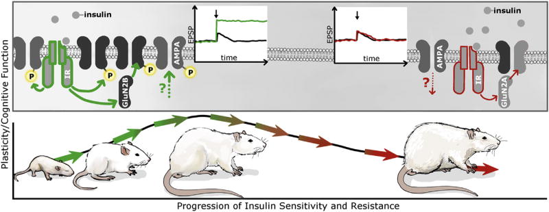
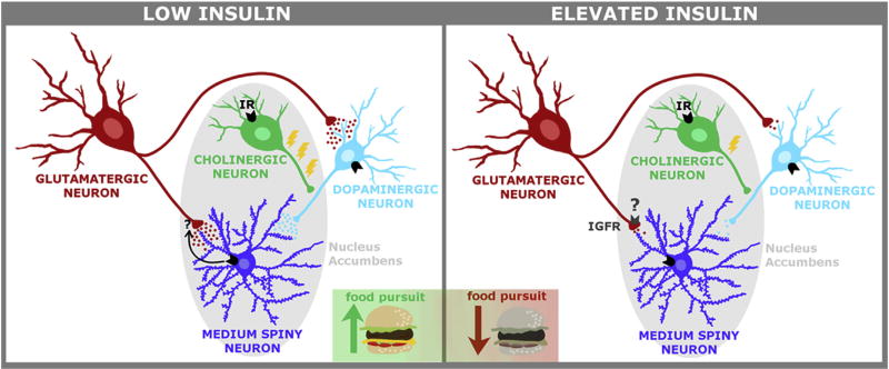
For decades the brain was erroneously considered an insulin insensitive organ. Although gaps in our knowledge base remain, conceptual frameworks are starting to emerge to provide insight into the mechanisms through which insulin facilitates critical brain functions like metabolism, cognition, and motivated behaviors. These diverse physiological and behavioral activities highlight the region-specific activities of insulin in the CNS; that is, there is an anatomical context to the activities of insulin in the CNS. Similarly, there is also a temporal context to the activities of insulin in the CNS. Indeed, brain insulin receptor activity can be conceptualized as a continuum in which insulin promotes neuroplasticity from development into adulthood where it is an integral part of healthy brain function. Unfortunately, brain insulin resistance likely contributes to neuroplasticity deficits in obesity and type 2 diabetes mellitus (T2DM). This neuroplasticity continuum can be conceptualized by the mechanisms through which insulin promotes cognitive function through its actions in brain regions like the hippocampus, as well as the ability of insulin to modulate motivated behaviors through actions in brain regions like the nucleus accumbens and the ventral tegmental area. Thus, the goals of this review are to highlight these anatomical, temporal, and functional contexts of insulin activity in these brain regions, and to identify potentially critical time points along this continuum where the transition from enhancement of neuroplasticity to impairment may take place. This article is part of the Special Issue entitled 'Metabolic Impairment as Risk Factors for Neurodegenerative Disorders.'
Keywords: Cognition; Glutamate; Hippocampus; Motivation; Nucleus accumbens; Ventral tegmental area.
Copyright © 2017 The Authors. Published by Elsevier Ltd.. All rights reserved.
Conflict of interest statement
The authors have no conflicts of interest to declare.
Figures
References
-
- Accili D, Drago J, Lee EJ, Johnson MD, Cool MH, Salvatore P, Asico LD, Jose PA, Taylor SI, Westphal H. Early neonatal death in mice homozygous for a null allele of the insulin receptor gene. Nat. Genet. 1996;12:106–109. - PubMed
-
- Banks WA. The source of cerebral insulin. Eur. J. Pharmacol. 2004;490:5–12. - PubMed
-
- Banks WA, DiPalma CR, Farrell CL. Impaired transport of leptin across the blood-brain barrier in obesity. Peptides. 1999;20:1341–1345. - PubMed
-
- Banks WA, Kastin AJ. Differential permeability of the blood-brain barrier to two pancreatic peptides: insulin and amylin. Peptides. 1998;19:883–889. - PubMed
Publication types
MeSH terms
Substances
Grants and funding
LinkOut - more resources
Full Text Sources
Other Literature Sources
Medical
