. 2018 Jan 11;145(1):dev154393.
doi: 10.1242/dev.154393.

Mapping the distribution of stem/progenitor cells across the mouse middle ear during homeostasis and inflammation

Affiliations

Mapping the distribution of stem/progenitor cells across the mouse middle ear during homeostasis and inflammation

Abigail S Tucker et al. Development. .

Abstract

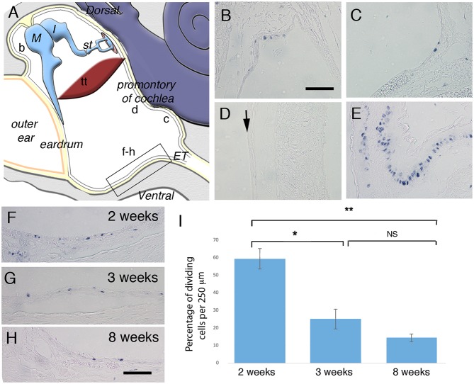
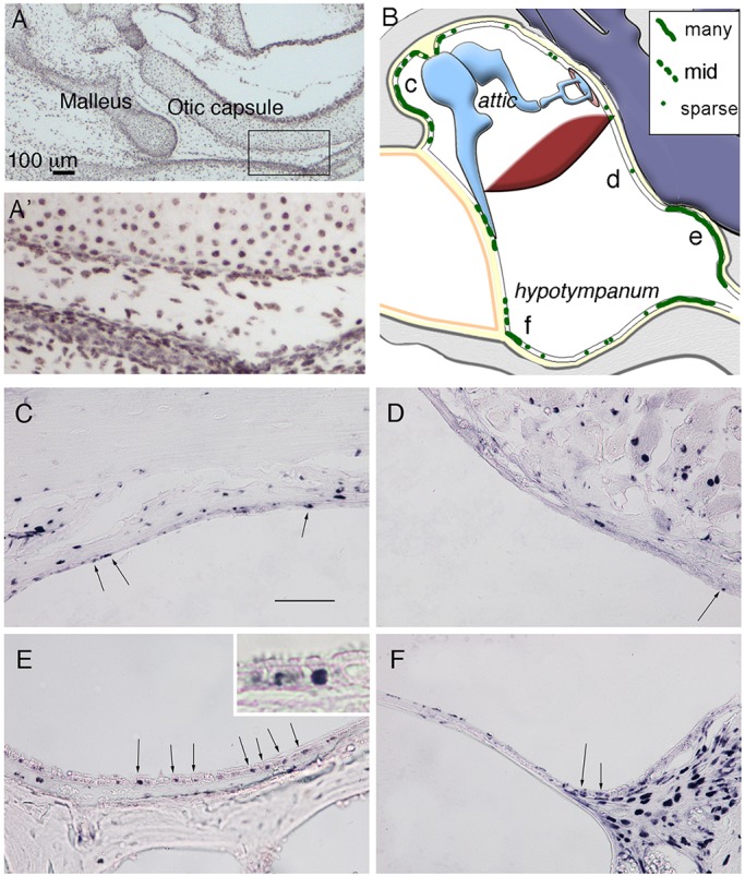
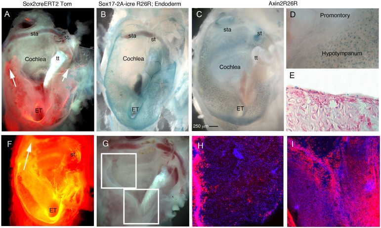
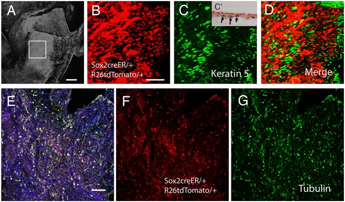
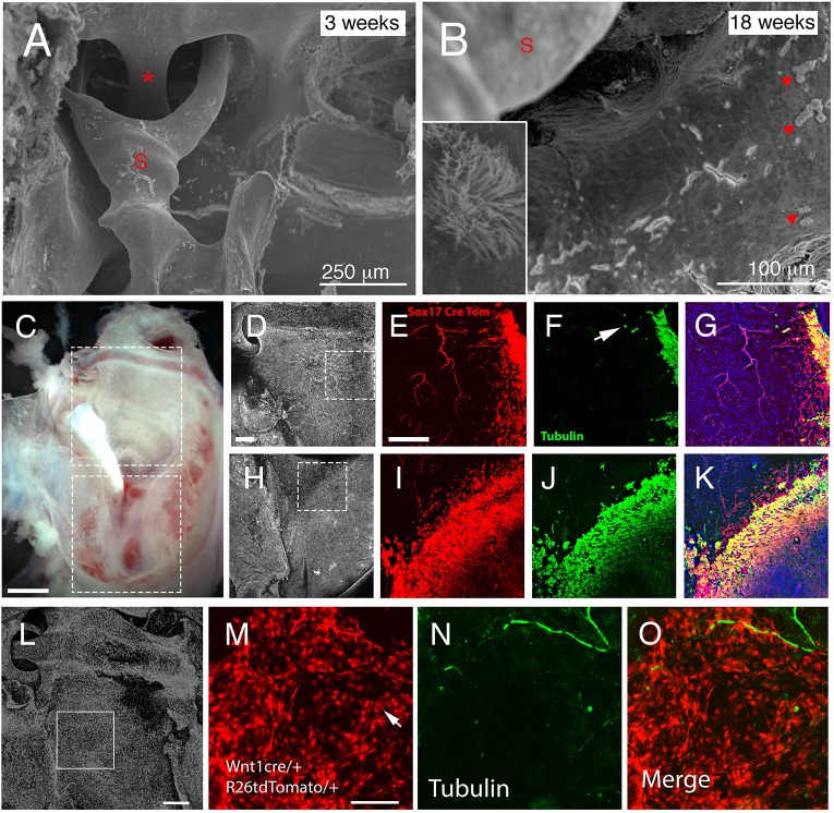
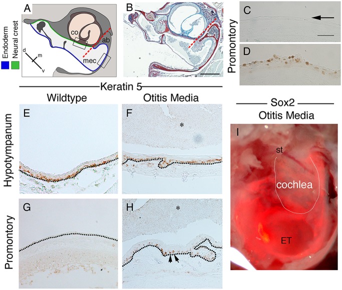
The middle ear epithelium is derived from neural crest and endoderm, which line distinct regions of the middle ear cavity. Here, we investigate the distribution of putative stem cell markers in the middle ear, combined with an analysis of the location of label-retaining cells (LRCs) to create a map of the middle ear mucosa. We show that proliferating cells and LRCs were associated with specific regions of the ear epithelium, concentrated in the hypotympanum at the base of the auditory bulla and around the ear drum. Sox2 was widely expressed in the endodermally derived ciliated pseudostratified epithelium of the hypotympanum. This part of the middle ear showed high levels of Wnt activity, as indicated by the expression of Axin2, a readout of Wnt signalling. Keratin 5 showed a more restricted expression within the basal cells of this region, with very little overlap between the Sox2- and keratin 5-positive epithelium, indicating that these genes mark distinct populations. Little expression of Sox2 or keratin 5 was observed in the neural crest-derived middle ear epithelium that lined the promontory, except in cases of otitis media when this epithelium underwent hyperplasia. This study lays the foundation for furthering our understanding of homeostasis and repair in the middle ear.

Keywords: Endoderm; Keratin 5; Label-retaining cells; Neural crest; Otitis media; Sox2.


Conflict of interest statement

Competing interestsThe authors declare no competing or financial interests.

Figures

Fig. 1.
Fig. 1.
Proliferation in distinct parts of the postnatal middle ear epithelium. (A) Schematic illustrating the middle ear cavity with b-d,f-h showing the position of B-D,F-H. The boxed region in A shows the area used for cell counts. (B-H) Frontal sections immunostained for PCNA. (B) Dorsal to eardrum. (C) Close to Eustachian tube. (D) Promontory over cochlea. Arrow indicates negative middle ear epithelium. (E) Proximal trachea epithelium. (F-H) Ventral epithelium showing the position of cell counts at 2 weeks (F), 3 weeks (G) and 8 weeks (H). (I) Graph showing average proliferating cell density in the hypotympanum (per 250 µm) at 2 (n=3 ears), 3 (n=3 ears) and 8 (n=3 ears) weeks. Error bars indicate s.e.m. *P<0.05, **P<0.005; NS, not significant. Student's unpaired t-test. M, malleus; I, Incus; st, stapes; tt, tensor tympani; ET, Eustachian tube. Scale bars: 100 µm in B-H.
Fig. 2.
Fig. 2.
Label-retaining cells are concentrated in specific parts of the middle ear. (A,A′) Immunostained frontal section of a P1 mouse middle ear, following treatment of BrdU in the mother’s drinking water for 7 days using an antibody against BrdU. All cells are labelled in the ear. The boxed region in A is enlarged in A′. (B) Schematic highlighting distribution of BrdU-positive cells (green) within an example 8-week-old postnatal middle ear. Letters c-f represent regions shown in C-F. (C-F) Frontal sections of 8-week-old postnatal middle ear immunostained against BrdU, in the epithelium of the attic close to the ear drum (C), overlying the cochlea (D), in the hypotympanum close to the Eustachian tube (E) and near the ear drum on the ventral side (F). Arrows in C-F indicate positive epithelial cells. Inset in E shows labeling in basal cells. Scale bars: 100 µm in A; 100 µm in C-F.
Fig. 3.
Fig. 3.
High levels of Sox2 expression and Wnt activity restricted to the endodermally derived part of the middle ear. (A-D) Whole-mount preparation of auditory bullae (AB) at P21 after ear drum removal showing the Eustachian tube (ET) ventrally (bottom) and stapes (st) dorsally (top). (E) Section of AB shown in C. (F-I) Whole-mount preparation of auditory bullae at 6 months. (A) Sox2creERTom mouse, tamoxifen injected 48 h prior to cull. Sox2-positive cells (and any progeny formed in the last 48 h) labelled in red. Arrows indicate tracts of red cells around the promontory. (B) Sox17-2A-icreR26R mouse, endoderm-derived cells and blood vessels stained blue. (C-E) Axin2R26R middle ears. Active Wnt signalling stains blue. (C) Whole bulla. (D) Positive scattered cells in hypotympanum near the ET, but not in the promontory overlying the cochlea. (E) Cryosection through the hypotympanum showing scattered blue cells in the epithelium, counterstained with Eosin. (F) AB; dark-field view showing Sox2-positive cells and progeny in orange. Tomato expression is also observed in the cochlea underlying the middle ear in stripes at 6 months. Arrow indicates Tomato-positive cells extending up to the attic around the promontory. (G) Dissected AB, boxed regions show areas highlighted by confocal microscopy in H,I. (H) Promontory and attic region with sparse isolated Tomato-positive cells. Large numbers of positive cells are observed at the margin of the promontory reaching up to the dorsal part of the middle ear. (I) Hypotympanum covered in Sox2-positive cells as at 2 days post-tamoxifen. The promontory (V-shape at top of image) only has a few positive cells in comparison. tt, tensor tympani muscle; st, stapes; ET, Eustachian tube; sta, stapedial artery running through stapes. Scale bar: 250 µm in A-C,F.
Fig. 4.
Fig. 4.
Keratin 5 and Sox2 mark distinct populations of cells in the middle ear epithelium. (A-G) Confocal images of the auditory bulla (AB). (A) DAPI image showing region of the hypotympanum imaged. Boxed area is enlarged in B-D. (B) Sox2creERTom mouse, tamoxifen injected 48 h prior to cull. (C) Keratin 5 immunostaining (green) of Sox2creERTom bulla. C′ shows a section through the hypotympanum with keratin 5-labelled cells in black (arrows). (D) Merged image of B and C showing only a small overlap between Sox2 and keratin 5. Cells expressing both show as yellow. (E-G) Sox2creERTom mouse tamoxifen injected 48 h prior to cull, labelled with acetylated α-tubulin to highlight ciliated cells. (E) Merged image; double-labelled cells are yellow. Blue indicates nuclei (DAPI). (F) Sox2creERTom (red). (G) Acetylated α-tubulin (green). There is a large overlap between these two markers. Scale bars: 200 µm in A; 50 µm in B-D; 100 µm in E-G.
Fig. 5.
Fig. 5.
Dual origin of ciliated cells in the ear. (A,B) Scanning electron microscopy images of the middle ear showing the attic region with the stapedial artery (asterisk) and stapes (S) inserting into the oval window in control mice. (A) In 3-week-old animals, no cilia were observed adjacent to the stapes. (B) In contrast, at 18 weeks, control mice displayed patches of ciliated cells (arrowheads). Inset shows magnification of the cilia patch. (C) Dissected auditory bulla of Sox17creERT/tdTom mouse. Boxes highlight region imaged by confocal microscopy. (D,H) DAPI staining highlighting nuclei. Boxes highlight the regions shown in E-G and I-K. (D-G) Lateral edges of attic. (H-K) Border of hypotympanum and promontory. (E,I) Some endoderm cells (red) appear to spread into the neural crest-derived regions of the attic and promontory. (F,J) The ciliated cells (acetylated α-tubulin, green) are mainly located overlapping with the endoderm but a few cells do not co-express the endoderm marker (arrow in F). (G,K) Merged image. Endoderm-derived ciliated cells are in yellow. (L-O) Dissected auditory bulla of Wnt1cre/tdTom mouse. DAPI staining highlighting nuclei in the attic region. Box highlights the region imaged in M-O. (M) Neural crest-derived epithelial cells show up as red. Arrow highlights a red cell. (N) Acetylated α-tubulin (green) highlights nerves and a few lone ciliated cells. (O) Merged image showing ciliated cell is of neural crest origin. Scale bars: 250 µm in A; 100 µm in B; 500 µm in C; 200 µm in D-K; 200 µm in L; 100 µm in M-O.
Fig. 6.
Fig. 6.
Changes to keratin 5, PCNA and Sox2 expression with otitis media. (A) Schematic of mouse middle ear in frontal section, highlighting the border between the endoderm and neural crest. (B) Trichrome stain of a Tbx1/+ middle ear with otitis media at 11.5 weeks. The ear is full of effusion and infiltrated cells, and the endodermally lined hypotympanum shows signs of hyperplasia. Dashed red lines in A,B indicate the border between neural crest and endoderm. (C-H) Frontal sections of mouse middle ear in Tbx1/+mice (D,F,H) and control littermates (C,E,G) at P28. (C,D) PCNA (brown) on promontory. Arrow in C highlights negative control epithelium. (E-H) Keratin 5 immunohistochemistry. In wild-type mice (E,G), keratin 5 is expressed by basal stem cells in the pseudostratified epithelium (E). (G) Only a few keratin 5-positive cells are observed in the simple epithelium overlying the cochlea. (F,H) In Tbx1/+ mice with otitis media, keratin 5 expression in the ventral middle ear looks similar to the wild type (F). (H) In contrast, overlying the cochlea, many keratin 5-positive cells were observed (arrows). (E-H) Dotted black lines indicate the basal lamina underlying the epithelium. (I) Whole-mount image of a Sox2creERT/tdTom auditory bulla with OM. Mouse injected 2 days prior to P21 to activate cre. Fluorescent image overlying bright-field image. Positive cells overlie the cochlea. st, stapes; ET, Eustachian tube. White line indicates the cochlea. Scale bars: 200 µm in B; in C, 100 µm for C-H.

References

    1. Andoniadou C. L., Matsushima D., Mousavy Gharavy S. N., Signore M., Mackintosh A. I., Schaeffer M., Gaston-Massuet C., Mollard P., Jacques T. S., Le Tissier P. et al. (2013). Sox2+ stem/progenitor cells in the adult mouse pituitary support organ homeostasis and have tumor-inducing potential. Cell Stem Cell 13, 433-445. 10.1016/j.stem.2013.07.004 - DOI - PubMed
    1. Arnold K., Sarkar A., Yram M. A., Polo J. M., Bronson R., Sengupta S., Seandel M., Geijsen N. and Hochedlinger K. (2011). Sox2+ adult stem and progenitor cells are important for tissue regeneration and survival of mice. Cell Stem Cell 9, 317-329. 10.1016/j.stem.2011.09.001 - DOI - PMC - PubMed
    1. Atef A. and Ayad E. E. (2004). Ciliary count in chronic suppurative otitis media: comparative quantitative study between mucosal and squamous types using scanning electron microscopy and image analysis. J. Laryngol. Otol. 118, 343-347. 10.1258/002221504323086516 - DOI - PubMed
    1. Danielian P. S., Muccino D. and Rowitch D. H. (1998). Modification of gene activity in mouse embryos in utero by a tamoxifen-inducible form of Cre recombinase. Curr. Biol. 8, 1323-1326. 10.1016/S0960-9822(07)00562-3 - DOI - PubMed
    1. Engert S., Liao W. P., Burtscher I. and Lickert H. (2009). Sox17-2A-iCre: a knock-in mouse line expressing Cre recombinase in endoderm and vascular endothelial cells. Genesis 47, 603-610. 10.1002/dvg.20540 - DOI - PubMed

Publication types