. 2017 Nov 15;9(11):4945-4953.
eCollection 2017.

Hsp90 inhibitor sensitizes TRAIL-mediated apoptosis via chop-dependent DR5 upregulation in colon cancer cells

Affiliations

Hsp90 inhibitor sensitizes TRAIL-mediated apoptosis via chop-dependent DR5 upregulation in colon cancer cells

Zhicheng Yao et al. Am J Transl Res. .

Abstract

Heat shock protein 90 (Hsp90), a molecular chaperone, is involved in a variety of physiological and pathological processes. Targeting Hsp90 by small molecules has been developed as an attractive strategy of anticancer therapy. In this study, we investigated the mechanism of Hsp90 inhibitor suppresses CRC growth and potentiates effects of other chemotherapeutic drugs. We found that Hsp90 inhibitor induces chop-dependent DR5 upregulation regardless of p53 status. Furthermore, DR5 is required for Hsp90 inhibitor-induced apoptosis. Hsp90 inhibitor also synergized with TRAIL to induce marked apoptosis via DR5 in CRC. Overall, our results illustrate DR5 play a key role in mediating the anticancer effects of Hsp90 inhibitor in CRC and suggest that DR5 expression can be used as an indicator of Hsp90 inhibitor sensitivity, which has important implications for it clinical applications.

Keywords: DR5; Hsp90 inhibitor; TRAIL; apoptosis; chop.


Conflict of interest statement

None.

Figures

Figure 1
Figure 1
17-AAG induces apoptosis in CRC. (A) The indicated cell lines were treated with increasing concentrations of 17-AAG for 72 hours. Cell proliferation was determined by MTS assay. (B) The indicated cell lines were treated with 17-AAG for 48 hours at indicated concentrations. Apoptosis was analyzed by Annexin V/PI staining followed by flow cytometry. (C) HCT116 cells were treated with 17-AAG with or without z-VAD-fmk, caspase 3/7 activity was determined by fluorogenic analysis. (D) HCT116 and RKO cells were treated with 1 μmol/L 17-AAG at indicated time point. Cleaved-caspase 3 and 8 were analyzed by western blotting. Results in (A-C) were expressed as means ± SD of three independent experiments. ***, P<0.001; **, P<0.01; *, P<0.05 compared to control cells.
Figure 2
Figure 2
Caspase 8 and FADD are required for Hsp90 inhibitor-induced apoptosis. (A) Parental and Cas 8-KD HCT116 cells were treated with 1 μmol/L 17-AAG for 48 hours. Apoptosis was analyzed by a nuclear fragmentation assay. (B) Parental and Cas 8-KD HCT116 cells were treated with 1 μmol/L 17-AAG for 48 hours. Cleaved-caspase 3 and 8 were analyzed by western blotting. (C) Parental and FADD-KD HCT116 cells were treated with 1 μmol/L 17-AAG for 48 hours. Lift, western blotting confirmed FADD depletion by shRNA. Right, apoptosis was analyzed by a nuclear fragmentation assay. (D) Parental and FADD-KD HCT116 cells were treated with 1 μmol/L 17-AAG for 48 hours. Cleaved-caspase 3 and 8 were analyzed by western blotting. Results in (A) and (C) were expressed as means ± SD of three independent experiments. ***, P<0.001; **, P<0.01 compared to control cells.
Figure 3
Figure 3
Hsp90 inhibitor induces DR5 induction in CRC. A. HCT116 cells were treated with 17-AAG for 24 hours at indicated concentration. DR5 mRNA expression was analyzed by RT-PCR. B. HCT116 cells were treated with 1 μmol/L 17-AAG for 24 hours. DR5 mRNA level was analyzed by gel electrophoresis. β-Actin was used as a control for normalization. C. HCT116 cells were treated with 1 μmol/L 17-AAG at indicated time point. Indicated protein was analyzed by western blotting. D. HCT116 cells were treated with 17-AAG for 24 hours at indicated concentration. DR5 was analyzed by western blotting. E. HCT116 cells were treated with 0.25 μmol/L 17-DMAG at indicated time point. DR5 was analyzed by western blotting.
Figure 4
Figure 4
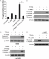
Hsp90 inhibitor sensitizes TRAIL-mediated apoptosis. A. HCT116 cells were treated with 1 μmol/L 17-AAG, 25 ng/mL TRAIL or their combination with or without 10 μmol/L z-VAD-fmk for 24 hours. Apoptosis was analyzed by a nuclear fragmentation assay. Results were expressed as means ± SD of three independent experiments. **, P<0.01 compared to control cells. B. HCT116 cells were treated with 1 μmol/L 17-AAG, 25 ng/mL TRAIL or their combination for 24 hours. Cleaved-caspase 3 and 8 were analyzed by western blotting. C. RKO or Lim2405 cells were treated with 1 μmol/L 17-AAG, 25 ng/mL TRAIL or their combination for 24 hours. Cleaved-caspase 3 was analyzed by western blotting. D. HCT116 cells were treated with the combination of 1 μmol/L 17-AAG and 25 ng/mL TRAIL with or without 10 μmol/Lz-VAD-fmk for 24 hours. Cleaved-caspase 3 was analyzed by western blotting.
Figure 5
Figure 5
DR5 is required for Hsp90 inhibitor-induced apoptosis. (A) Parental and DR5-KD HCT116 cells were treated with 1 μmol/L 17-AAG for 24 hours. DR5 was analyzed by western blotting. (B) Parental and DR5-K DHCT116 cells were treated with 1 μmol/L 17-AAG for 48 hours. Apoptosis was analyzed by a nuclear fragmentation assay. (C) Parental and DR5-KD HCT116 cells were treated with 1 μmol/L 17-AAG for 24 hours. Cleaved-caspase 3 was analyzed by western blotting. (D) Parental and DR5-KD HCT116 cells were treated with 1 μmol/L 17-AAG, 25 ng/mL TRAIL or their combination for 24 hours. Apoptosis was analyzed by a nuclear fragmentation assay. (E) Parental and DR5-KD HCT116 cells were treated with the combination of 1 μmol/L 17-AAG and 25 ng/mL TRAIL with or without 10 μmol/Lz-VAD-fmk for 24 hours. Cleaved-caspase 3 and 8 were analyzed by western blotting. (F) RKO cells were transfect with sicontrol or siDR5 for 24 hours, and then treated with the combination of 1 μmol/L 17-AAG and 25 ng/mL TRAIL with or without 10 μmol/L z-VAD-fmk for 24 hours. Cleaved-caspase 3 and 8 were analyzed by western blotting. Results in (B) and (D) were expressed as means ± SD of three independent experiments. ***, P<0.001; **, P<0.01; *, P<0.05 compared to control cells.
Figure 6
Figure 6
Hsp90 inhibitor induces chop-mediated DR5 expression. A. HCT116 cells were treated with 1 μmol/L 17-AAG at indicated time point, phospho-p65, p65 and MZF1 were analyzed by western blotting. B. HCT116 cells were transfect with si control or sichop for 24 hours, and then treated with 1 μmol/L 17-AAG for 48 hours. Chop, DR5 and cleaved-caspase 3 were analyzed by western blotting. C. HCT116 cells were transfected with si control or sichop for 24 hours, and then treated with 1 μmol/L 17-AAG for 48 hours. Apoptosis was analyzed by a nuclear fragmentation assay. Results were expressed as means ± SD of three independent experiments. **, P<0.01 compared to control cells. D. HCT116 cells were treated with 1 μmol/L 17-AAG at indicated time point. Chop and Bip were analyzed by western blotting. E. HCT116 cells were treated with 1 μmol/L 17-AAG at indicated time point. ChopmRNA level was analyzed by gel electrophoresis. β-Actin was used as a control for normalization. F. HCT116 cells were treated with 0.25 μmol/L 17-DMAG at indicated time point. Chop was analyzed by western blotting. G. Chromatin immunoprecipitation (ChIP) was performed using anti-chop antibody on HCT116 cells following 17-AAG treatment for 12 hours. ChIP with the control IgG was used as a control. PCR was carried out using primers surrounding the chop binding sites in the DR5 promoter.

Similar articles

Cited by

References

    1. Siegel RL, Miller KD, Jemal A. Cancer statistics, 2016. CA Cancer J Clin. 2016;66:7–30. - PubMed
    1. Goldberg RM. Therapy for metastatic colorectal cancer. Oncologist. 2006;11:981–987. - PubMed
    1. Chua CS, Low H, Sim TS. Co-chaperones of Hsp90 in Plasmodium falciparum and their concerted roles in cellular regulation. Parasitology. 2014;141:1177–1191. - PubMed
    1. Chehab M, Caza T, Skotnicki K, Landas S, Bratslavsky G, Mollapour M, Bourboulia D. Targeting Hsp90 in urothelial carcinoma. Oncotarget. 2015;6:8454–8473. - PMC - PubMed
    1. Sidera K, Patsavoudi E. HSP90 inhibitors: current development and potential in cancer therapy. Recent Pat Anticancer Drug Discov. 2014;9:1–20. - PubMed

LinkOut - more resources