. 2017 Nov 15;9(11):5094-5104.
eCollection 2017.

Scutellarin suppresses human colorectal cancer metastasis and angiogenesis by targeting ephrinb2

Affiliations

Scutellarin suppresses human colorectal cancer metastasis and angiogenesis by targeting ephrinb2

Ping Ting Zhu et al. Am J Transl Res. .

Abstract

Tumor induced angiogenesis is an attractive target for anti-cancer drug treatment. Scutellarin, which is a native compound derived from scutellaria altissima leaves, has already been proved to possess anti-tumor activities. Nevertheless, their effects in colorectal cancer metastasis and angiogenesis have not been evaluated. In order to reveal the anti-angiogenic and anti-metastasis capacity of scutellarin, wound healing and Transwell chamber inserts invasion were done in colorectal cancer cells, and cell proliferation as wells colony formation were conducted to identify the proliferation inhibition of colorectal cancer in vitro. The growth inhibition of scutellarin was further definite by a mouse colorectal xenograft model in vivo. Herein, we demonstrated scutellarin suppressed colorectal cancer cell viability and colony formation in vitro, and remarkably reduced tumor growth in vivo mouse xenografts. Additionally, scutellarin restrained colorectal cancer cells-induced angiogenesis, inhibited human umbilical vascular endothelial cells (HUVECs) migration, tube formation of HUVECs, and micro-vessel formation in chick embnyo chorioallantoic menbreme (CAM) assay. Altogether, our results exhibited the evidence that scutellarin inhibit colorectal cancer angiogenesis and metastasis via targeting ephrinb2 signaling, with the potential of an anti-tumor agent for cancer treatment.

Keywords: Colorectal cancer; angiogenesis; ephrinb2; metastasis; scutellarin.


Conflict of interest statement

None.

Figures

Figure 1
Figure 1
Scutellarin inhibits CRC cell growth and mobility. A. HT29, HCT116, LOVO and SW480 cells were exposed with scutellarin, and cell viability were measured. Data are the mean ± SD. B. Scutellarin inhibited colony formation of CRC cells. One day after pre-treatment various concentrations of scutellarin, cells were plated into 6-well plate cultured for 14 days and cells colonies were counted. Data represented the mean ± SD. *p < 0.05, **p < 0.01 compared to control. C. Transwell analysis was conducted to determine the invasion of CRC cells. Cells invaded the Transwell membrane were counted. *p < 0.05, **p < 0.01 compared to control. D. Cells were pre-treated with scutellarin for 24 h, the wound were created and percentage of wound closure over 24 h was measured. *p < 0.05, **p < 0.01 compared to control.
Figure 2
Figure 2
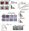
Effect of scutellarin on tumor growth and angiogenesis in vivo. A. Treatment with scutellarin was initiated when tumor volumes reached 100 mm3. Tumor weights were measured at the end of the experiment. B. 1 × 10⁶ HCT116 cells were s.c. injected and nude mice were oral administrated with the scutellarin. Xenograft tumor volumes were measured three times a week by a caliper. C. Immunohistochemical analysis showed that scutellarin inhibited Ki67 positive cells in colorectal cancer xenografts (upper panel). Immunohistochemical analysis showed that scutellarin inhibited CD34-positive blood vessels in colorectal cancer xenografts (lower panel). *p < 0.05, **p < 0.01 vs. vehicle. D. Representative picture of the lungs from mice injected intravenously with HCT116 cells after treatment with vehicle or 10 mg/kg scutellarin. *p < 0.05, compared to vehicle.
Figure 3
Figure 3
Effect of scutellarin on angiogenesis and tube formation. A. Effect of scutellarin on the angiogenesis in CAM. In the control group, the blood vessels grew normally. Treated with various concentrations of scutellarin, CAM blood vessels were significantly inhibited. B. Effect of scutellarin on the tube formation. In the control group, HUVECs cells formed the tube structure on the Matrigel. In the scutellarin treated group, tube foramtion was inhibited in dose-dependent manner. Data are expressed as means ± SD. *p < 0.05, **p < 0.01, compared to the control group. C. HUVECs were plated to six-well plate. A scratch was created and cells were treated with CM of HCT116 cells treated or un-treated with scutellarin. The percentage of wound clousre was quantified. D. Scutellarin inhibited the invasion of HUVECs induced by the CM of HCT116 cells treated or un-treated with scutellarin. The results shown were representative of three independent experiments. *p < 0.05; **p < 0.01, compare to control.
Figure 4
Figure 4
Effects of scutellarin on the expression of cells metastasis related genes. A. 26 genes involved in the migration and invasion processes were screened by PCR array. HCT116 cells were treated or un-treated with scutellarin for 48 h. RNA extraction and retrotranscription, the resulting cDNA was applied in the PCR array and amplified. Data are reported as fold compared to untreated HCT116 cells. B. Immunoblotting assay of cell lysates from HCT116 cells treated or untreated with scutellarin. C. HCT116 cells were transfected with constitutively active form of ephrinb2 and the expression of ephrinb2 was determined by western blot. D. In the presence of scutellarin, wound healing assay was subjected to assess the cell motility after ephrinb2 over-expression. Data are expressed as means ± SD. **p <0.01 compared to control cells, ##p < 0.01 compared to cells transfected with vector. E. HUVECs cells were performed to wound closure analysis in the absence of CM from HCT116 cells treated or un-treated with scutellarin. F. Tube formation was performed in the absence of CM from HCT116 cells treated or un-treated with scutellarin. Columns are data collected from three independent experiments and are average ± SD. values. **p <0.01 compared to control cells, ##p < 0.01 compared to cells transfected with vector.

References

    1. Huang Y, Duan H, Sun Y. Elevated expression of SAG/ROC2/Rbx2/Hrt2 in human colon carcinomas: SAG does not induce neoplastic transformation, but antisense SAG transfection inhibits tumor cell growth. Mol Carcinog. 2001;30:62–70. - PubMed
    1. Zhang W, Shi X, Peng Y, Wu M, Zhang P, Xie R, Wu Y, Yan Q, Liu S, Wang J. HIF-1alpha promotes epithelial-mesenchymal transition and metastasis through direct regulation of ZEB1 in colorectal cancer. PLoS One. 2015;10:e0129603. - PMC - PubMed
    1. Qin Y, Tang B, Hu CJ, Xiao YF, Xie R, Yong X, Wu YY, Dong H, Yang SM. An hTERT/ZEB1 complex directly regulates E-cadherin to promote epithelial-to-mesenchymal transition (EMT) in colorectal cancer. Oncotarget. 2016;7:351–361. - PMC - PubMed
    1. Srinivasainagendra V, Sandel MW, Singh B, Sundaresan A, Mooga VP, Bajpai P, Tiwari HK, Singh KK. Migration of mitochondrial DNA in the nuclear genome of colorectal adenocarcinoma. Genome Med. 2017;9:31. - PMC - PubMed
    1. Chan JY, Tan BK, Lee SC. Scutellarin sensitizes drug-evoked colon cancer cell apoptosis through enhanced caspase-6 activation. Anticancer Res. 2009;29:3043–3047. - PubMed

LinkOut - more resources