

Downregulation of cathepsin G reduces the activation of CD4+ T cells in murine autoimmune diabetes
- PMID: 29218110
- PMCID: PMC5714796
Downregulation of cathepsin G reduces the activation of CD4+ T cells in murine autoimmune diabetes
Abstract
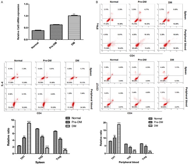
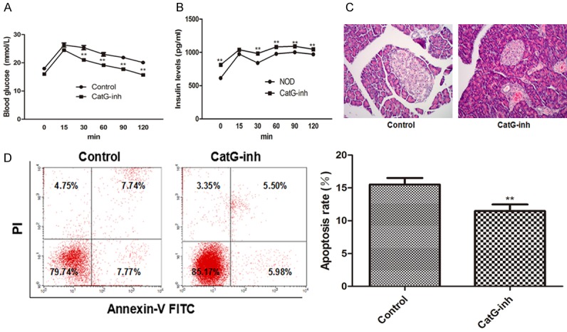
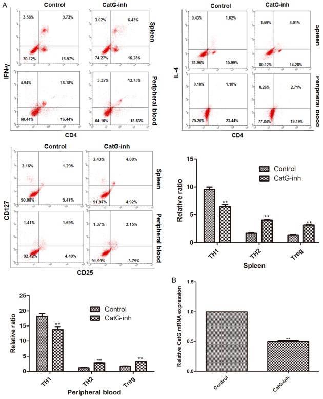
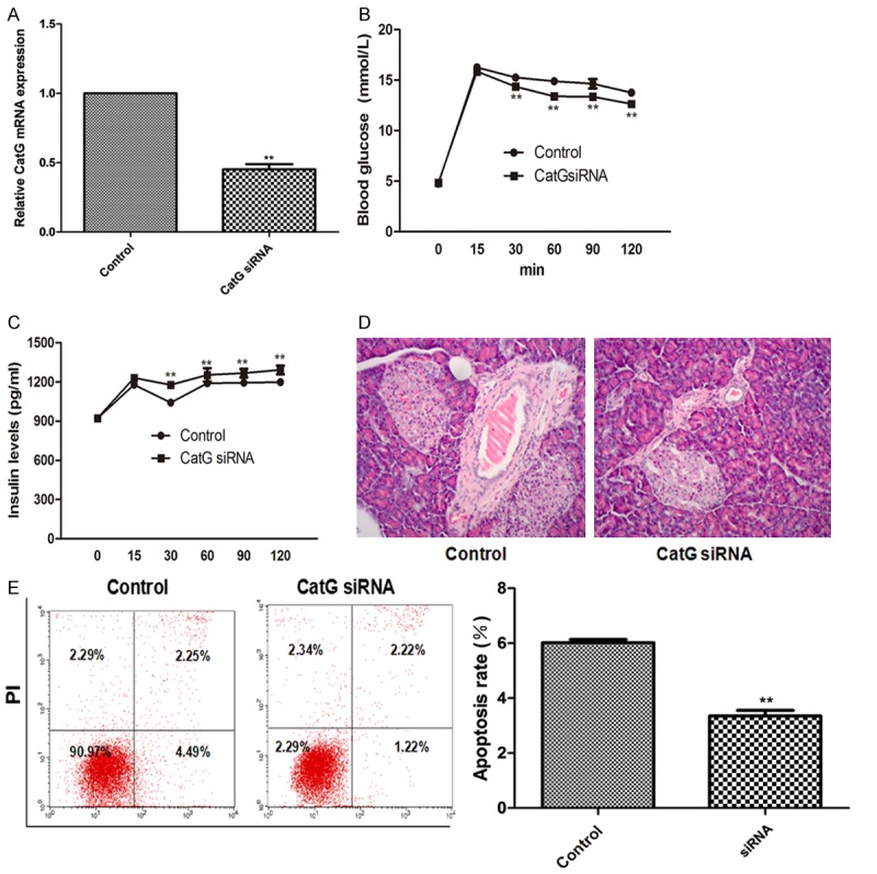
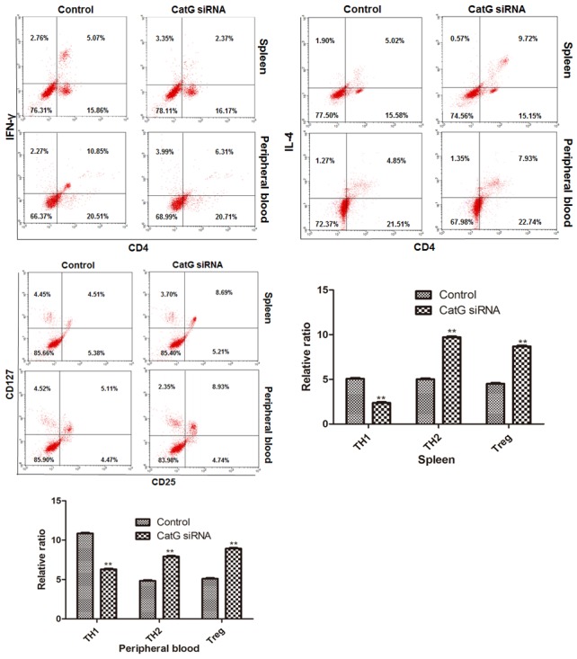
Type 1 diabetes mellitus (T1DM) is an autoimmune disease due to progressive injury of islet cells mediated by T lymphocytes (T cells). Our previous studies have shown that only cathepsin G (CatG), not other proteases, is involved in the antigen presentation of proinsulin, and if the presentation is inhibited, the activation of CD4+ T cells induced by proinsulin is alleviated in T1DM patients, and CatG-specific inhibitor reduces the activation of CD4+ cells induced by proinsulin in T1DM patients. Therefore, we hypothesize that CatG may play an important role in the activation of CD4+ T cells in T1DM. To this end, mouse studies were conducted to demonstrate that CatG impacts the activation of CD4+ T cells in non-obese diabetic (NOD) mice. CatG gene expression and the activation of CD4+ T cells were examined in NOD mice. The effect of CatG inhibitor was investigated in NOD mice on the activation of CD4+ T cells, islet β cell function, islet inflammation and β-cell apoptosis. Furthermore, NOD mice were injected with CatG siRNA in early stage to observe the effect of CatG knockdown on the activation status of CD4+ T cells and the progression of diabetes. During the pathogenesis of diabetes, the expression level of CatG in NOD mice gradually increased and the CD4+ T cells were gradually activated, resulting in more TH1 cells and less TH2 and Treg cells. Treatment with CatG-specific inhibitor reduced the blood glucose level, improved the function of islet β cells and reduced the activation of CD4+ T cells. Early application of CatG siRNA improved the function of islet β cells, reduced islet inflammation and β cell apoptosis, and lowered the activation level of CD4+ T cells, thus slowing down the progression of diabetes.
Keywords: CD4+ T cells; NOD mice; cathepsin G; type 1 diabetes mellitus.
Conflict of interest statement
None.
Figures
Similar articles
-
Vitamin D supplementation induces CatG-mediated CD4+ T cell inactivation and restores pancreatic β-cell function in mice with type 1 diabetes.Am J Physiol Endocrinol Metab. 2022 Jan 1;322(1):E74-E84. doi: 10.1152/ajpendo.00066.2021. Epub 2021 Nov 15. Am J Physiol Endocrinol Metab. 2022. PMID: 34779254
-
In vitro xenorecognition of adult pig pancreatic islet cells by splenocytes from nonobese diabetic or non-diabetes-prone mice.Transplantation. 1998 Sep 15;66(5):633-8. doi: 10.1097/00007890-199809150-00015. Transplantation. 1998. PMID: 9753345
-
Vaccination with a co-expression DNA plasmid containing GAD65 fragment gene and IL-10 gene induces regulatory CD4(+) T cells that prevent experimental autoimmune diabetes.Diabetes Metab Res Rev. 2016 Sep;32(6):522-33. doi: 10.1002/dmrr.2780. Epub 2016 Mar 8. Diabetes Metab Res Rev. 2016. PMID: 26797873
-
Cellular and molecular roles of beta cell autoantigens, macrophages and T cells in the pathogenesis of autoimmune diabetes.Arch Pharm Res. 1999 Oct;22(5):437-47. doi: 10.1007/BF02979150. Arch Pharm Res. 1999. PMID: 10549569 Review.
-
Control of type 1 autoimmune diabetes by naturally occurring CD4+CD25+ regulatory T lymphocytes in neonatal NOD mice.Ann N Y Acad Sci. 2005 Jun;1051:72-87. doi: 10.1196/annals.1361.048. Ann N Y Acad Sci. 2005. PMID: 16126946 Review.
Cited by
-
Hindrance of the Proteolytic Activity of Neutrophil-Derived Serine Proteases by Serine Protease Inhibitors as a Management of Cardiovascular Diseases and Chronic Inflammation.Front Chem. 2021 Nov 15;9:784003. doi: 10.3389/fchem.2021.784003. eCollection 2021. Front Chem. 2021. PMID: 34869231 Free PMC article. Review.
-
Two to Tango: Dialogue between Adaptive and Innate Immunity in Type 1 Diabetes.J Diabetes Res. 2020 Jul 30;2020:4106518. doi: 10.1155/2020/4106518. eCollection 2020. J Diabetes Res. 2020. PMID: 32802890 Free PMC article. Review.
-
The duplexity of insulin: The integrated bioinformatics analysis and machine learning identified key genes for type 2 diabetes.Biochem Biophys Rep. 2025 Jun 24;43:102099. doi: 10.1016/j.bbrep.2025.102099. eCollection 2025 Sep. Biochem Biophys Rep. 2025. PMID: 40641746 Free PMC article.
-
Progress in the Relationship between Vitamin D Deficiency and the Incidence of Type 1 Diabetes Mellitus in Children.J Diabetes Res. 2022 Sep 2;2022:5953562. doi: 10.1155/2022/5953562. eCollection 2022. J Diabetes Res. 2022. PMID: 36090587 Free PMC article. Review.
-
Cathepsin G and Its Role in Inflammation and Autoimmune Diseases.Arch Rheumatol. 2018 Jan 22;33(4):498-504. doi: 10.5606/ArchRheumatol.2018.6595. eCollection 2018 Dec. Arch Rheumatol. 2018. PMID: 30874236 Free PMC article. Review.
References
-
- Eisenbarth GS. Type I diabetes mellitus. A chronic autoimmune disease. N Engl J Med. 1986;314:1360–1368. - PubMed
-
- Roep BO, Tree TI. Immune modulation in humans: implications for type 1 diabetes mellitus. Nat Rev Endocrinol. 2014;10:229–242. - PubMed
-
- Lernmark A, Larsson HE. Immune therapy in type 1 diabetes mellitus. Nat Rev Endocrinol. 2013;9:92–103. - PubMed
-
- Moran A, Bundy B, Becker DJ, DiMeglio LA, Gitelman SE, Goland R, Greenbaum CJ, Herold KC, Marks JB, Raskin P, Sanda S, Schatz D, Wherrett DK, Wilson DM, Krischer JP, Skyler JS Type 1 Diabetes TrialNet Canakinumab Study Group; Pickersgill L, de Koning E, Ziegler AG, Boehm B, Badenhoop K, Schloot N, Bak JF, Pozzilli P, Mauricio D, Donath MY, Castano L, Wagner A, Lervang HH, Perrild H, Mandrup-Poulsen T AIDA Study Group. Interleukin-1 antagonism in type 1 diabetes of recent onset: two multicentre, randomised, double-blind, placebo-controlled trials. Lancet. 2013;381:1905–1915. - PMC - PubMed
-
- Ludvigsson J, Krisky D, Casas R, Battelino T, Castano L, Greening J, Kordonouri O, Otonkoski T, Pozzilli P, Robert JJ, Veeze HJ, Palmer J, Samuelsson U, Elding Larsson H, Aman J, Kardell G, Neiderud Helsingborg J, Lundstrom G, Albinsson E, Carlsson A, Nordvall M, Fors H, Arvidsson CG, Edvardson S, Hanas R, Larsson K, Rathsman B, Forsgren H, Desaix H, Forsander G, Nilsson NO, Akesson CG, Keskinen P, Veijola R, Talvitie T, Raile K, Kapellen T, Burger W, Neu A, Engelsberger I, Heidtmann B, Bechtold S, Leslie D, Chiarelli F, Cicognani A, Chiumello G, Cerutti F, Zuccotti GV, Gomez Gila A, Rica I, Barrio R, Clemente M, Lopez Garcia MJ, Rodriguez M, Gonzalez I, Lopez JP, Oyarzabal M, Reeser HM, Nuboer R, Stouthart P, Bratina N, Bratanic N, de Kerdanet M, Weill J, Ser N, Barat P, Bertrand AM, Carel JC, Reynaud R, Coutant R, Baron S. GAD65 antigen therapy in recently diagnosed type 1 diabetes mellitus. N Engl J Med. 2012;366:433–442. - PubMed
LinkOut - more resources
Full Text Sources
Research Materials