

Review
doi: 10.1200/JCO.2017.75.3467.
Epub 2017 Dec 8.
Biology and Management of Undifferentiated Pleomorphic Sarcoma, Myxofibrosarcoma, and Malignant Peripheral Nerve Sheath Tumors: State of the Art and Perspectives
Affiliations
- PMID: 29220302
- PMCID: PMC5759316
- DOI: 10.1200/JCO.2017.75.3467
Item in Clipboard
Review
Biology and Management of Undifferentiated Pleomorphic Sarcoma, Myxofibrosarcoma, and Malignant Peripheral Nerve Sheath Tumors: State of the Art and Perspectives
J Clin Oncol.
.
Abstract
Undifferentiated pleomorphic sarcomas, myxofibrosarcomas, and malignant peripheral nerve sheath tumors are characterized by complex genomic characteristics and aggressive clinical behavior. Recent advances in the understanding of the pathogenesis of these tumors may allow for the development of more-effective innovative therapeutic strategies, including immunotherapies. This review describes the current knowledge of the epidemiology, clinical presentation, treatment, and pathogenesis of these tumors and highlights ongoing and future research.
Figures
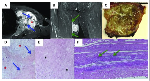
Magnetic resonance imaging (MRI) and histologic features of myxofibrosarcoma (MFS). (A) MFS of the left-side axillary region. Sagittal T2-weighted MRI shows three components: myxoid (red asterisk), fibrotic (blue arrow), and necrotic (black asterisk). (B) MFS of the right thigh. Axial T1-weighted MRI shows tumor spread along the fascial plane (green arrow, tail sign). (C) Macroscopic features of MFS. (D to F) Grossly, multinodular growth pattern and gelatinous, myxoid cut surface are shown. Microscopic features of MFS show the presence of three components: myxoid (red asterisk), fibrotic (arrow), and necrotic (black asterisk).
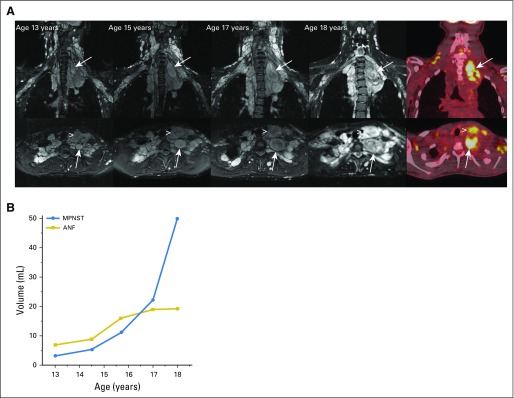
MRI and metabolic features of neurofibromas and malignant peripheral nerve sheath tumors (MPNSTs). (A) Axial (top panel) and coronal (bottom panel) short T1 inversion recovery magnetic resonance imaging of neck and chest plexiform neurofibroma in a female teenager with neurofibromatosis type 1. Neck pain for several months was attributed to stress and a heavy backpack. Development and progressive enlargement of a distinct nodular lesion were found in the neck and upper chest (arrows). In addition, growth of an anterior neck nodular lesion was found (arrowhead). [18F]fluorodeoxyglucose positron emission tomography at age 18 years demonstrated [18F]fluorodeoxyglucose avidity of the two nodular lesions. Biopsy of the deep nodular lesion (arrow) at age 18 years confirmed intermediate-grade MPNST. Surgical resection of MPNST and the anterior neck nodular lesion confirmed atypical neurofibroma (ANF). (B) Volume measurements of the deep and anterior neck nodular lesions over time showed parallel lesion growth from ages 13 to 15 years. Accelerated growth of the deep nodular lesion subsequent to this is concerning for malignant transformation.
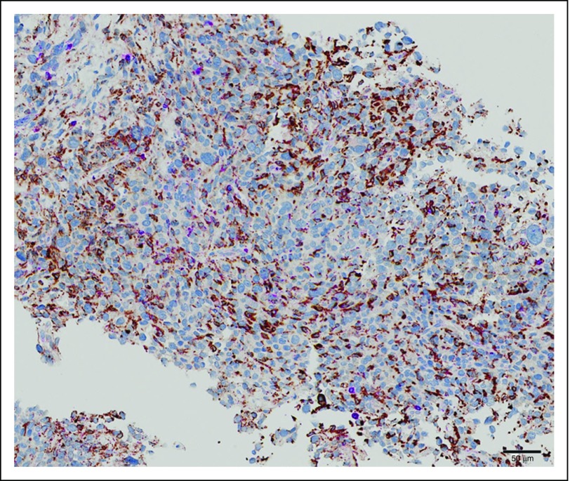
Massive infiltration by CD63/CD168+ macrophages in undifferentiated pleomorphic sarcoma.
References
-
- O’Brien JE, Stout AP: Malignant fibrous xanthomas. Cancer 17:1445-1455, 1964 - PubMed
-
- Le Doussal V, Coindre JM, Leroux A, et al. : Prognostic factors for patients with localized primary malignant fibrous histiocytoma: A multicenter study of 216 patients with multivariate analysis. Cancer 77:1823-1830, 1996 - PubMed
-
- Coindre JM, Mariani O, Chibon F, et al. : Most malignant fibrous histiocytomas developed in the retroperitoneum are dedifferentiated liposarcomas: A review of 25 cases initially diagnosed as malignant fibrous histiocytoma. Mod Pathol 16:256-262, 2003 - PubMed
-
- Fletcher CD: Pleomorphic malignant fibrous histiocytoma: Fact or fiction? A critical reappraisal based on 159 tumors diagnosed as pleomorphic sarcoma. Am J Surg Pathol 16:213-228, 1992 - PubMed
-
- Fletcher CD, Gustafson P, Rydholm A, et al. : Clinicopathologic re-evaluation of 100 malignant fibrous histiocytomas: Prognostic relevance of subclassification. J Clin Oncol 19:3045-3050, 2001 - PubMed
Publication types
MeSH terms
Substances
LinkOut - more resources
Full Text Sources
Other Literature Sources
Medical
