. 2017 Dec 8;12(12):e0188989.
doi: 10.1371/journal.pone.0188989. eCollection 2017.

Substitutions of PrP N-terminal histidine residues modulate scrapie disease pathogenesis and incubation time in transgenic mice

Affiliations

Substitutions of PrP N-terminal histidine residues modulate scrapie disease pathogenesis and incubation time in transgenic mice

Sabina Eigenbrod et al. PLoS One. .

Abstract

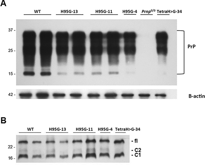
Prion diseases have been linked to impaired copper homeostasis and copper induced-oxidative damage to the brain. Divalent metal ions, such as Cu2+ and Zn2+, bind to cellular prion protein (PrPC) at octapeptide repeat (OR) and non-OR sites within the N-terminal half of the protein but information on the impact of such binding on conversion to the misfolded isoform often derives from studies using either OR and non-OR peptides or bacterially-expressed recombinant PrP. Here we created new transgenic mouse lines expressing PrP with disrupted copper binding sites within all four histidine-containing OR's (sites 1-4, H60G, H68G, H76G, H84G, "TetraH>G" allele) or at site 5 (composed of residues His-95 and His-110; "H95G" allele) and monitored the formation of misfolded PrP in vivo. Novel transgenic mice expressing PrP(TetraH>G) at levels comparable to wild-type (wt) controls were susceptible to mouse-adapted scrapie strain RML but showed significantly prolonged incubation times. In contrast, amino acid replacement at residue 95 accelerated disease progression in corresponding PrP(H95G) mice. Neuropathological lesions in terminally ill transgenic mice were similar to scrapie-infected wt controls, but less severe. The pattern of PrPSc deposition, however, was not synaptic as seen in wt animals, but instead dense globular plaque-like accumulations of PrPSc in TgPrP(TetraH>G) mice and diffuse PrPSc deposition in (TgPrP(H95G) mice), were observed throughout all brain sections. We conclude that OR and site 5 histidine substitutions have divergent phenotypic impacts and that cis interactions between the OR region and the site 5 region modulate pathogenic outcomes by affecting the PrP globular domain.


Conflict of interest statement

Competing Interests: The authors have read the journal's policy and have the following conflicts: after finalizing the experiments, three of the authors left the ZNP and are currently employed by commercial companies: Servier Deutschland GmbH (SE), MFD Diagnostic GmbH (MM), and BASF (NP). This does not alter the authors' adherence to all the PLOS ONE policies on sharing data and materials.

Figures

Fig 1
Fig 1. PrPC levels and glycosylation profile in healthy wild-type and transgenic mice expressing mutant PrP.
(A) Brain homogenates (10 μg per lane) derived from mice expressing full length mouse wild-type (wt), PrP(H95G) lines 4, 11 and 13, PrP(TetraH>G) line 34 and PrP null controls (Prnp0/0) were subjected to immunoblot analysis using monoclonal antibody SHA31. Blots were reprobed for β-actin to control for equal loading. Molecular weight is indicated on the left (in kDa). (B) Removal of N-linked glycans on PrPC encoded by the PrP(H95G), lines 4, 11, and 13 as well as PrP(TetraH>G), line 34 using peptide N-glycosidase F (PNGase F) and probing of the treated samples with monoclonal antibody SHA31. fl = full-length PrP.
Fig 2
Fig 2. Neuropathological changes and PrPSc distribution in hippocampal and cerebellar sections of prion-challenged mice.
(A) First column: Paraffin-embedded tissue (PET) blots demonstrating PrPSc deposits in hippocampus and cerebellum of RML-infected wild-type (C57/129Sv) and PrP(TetraH>G), line 34 mice. There are plaque-like deposits in and adjacent to the corpus callosum and the internal granular cell layer of the cerebellum in PrP(TetraH>G) mice. Second column: Corresponding PrPSc immunhistochemistry (upper field: hippocampus and corpus callosum; lower field: cerebellum; monoclonal antibody CDC1; scale bar: 50 μm). Third column: Hematoxylin and eosin stainings (H&E) of hippocampal/corpus callosum and cerebellar sections demonstrating spongiform changes (scale bars of hippocampus/corpus callosum: 100 μm; of cerebellum: 200 μm). Fourth column: GFAP immunostaining demonstrating gliosis (scale bars as in third column). (B) Corresponding histopathological and immunohistochemical examination of hippocampal brain sections derived from terminally ill PrPH95G mice and corresponding wt controls (scale bars: 200 μm). Three independent Tg lines (lines 4, 11 and 13) were assessed for these analyses.
Fig 3
Fig 3. Lesion profiles induced by mouse-adapted scrapie isolate RML in wild-type and transgenic mice expressing mutant PrP.
The extent of spongiform change (A) and reactive gliosis (C) in brain sections of terminally ill wt and PrP(TetraH>G) was assessed semi-quantitatively in a blinded fashion in nine areas of grey matter and three areas of white matter by lesion profiling. Animals were scored on a scale of 0–5 in each specific area, and mean scores (n = 6 (C57/129Sv), versus n = 7 (PrP(TetraH>G), line 34), respectively) are shown graphically (error bars plus SD). Blue diamonds: C57/129Sv. Red squares: PrP(TetraH>G). Analogous data from PrP(H95G) mice are shown in panels B and D (n = 4 (PrP(H95G), line 13), n = 5 (PrP(H95G), line 4) and n = 7 (PrP(H95G), line 11), respectively); data for C57/129Sv wt animals has been re-plotted for comparative purposes. Blue diamonds: C57/129Sv. Red squares: PrP(H95G), line 4. Green triangles: PrP(H95G), line 11. Grey circles: PrP(H95G), line 13. Scoring areas as follows: Grey matter: 1, dorsal medulla, 2, cerebellar cortex, 3, superior colliculus, 4, hypothalamus, 5, medial thalamus, 6, hippocampus, 7, septum, 8, medial cerebral cortex at septum level, 9, medial cerebral cortex at thalamus level. White matter: 1*, cerebellar white matter, 2*, mesencephalic tegmentum, 3* pyramidal tract.
Fig 4
Fig 4. Western blot analysis of brain lysates from RML-infected wt and transgenic mice for total PrPC and PrPSc using monoclonal antibody 4H11.
(A) Brain homogenates from terminally ill wt and PrP(TetraH>G) line 34 mice were either left untreated (- PK) or subjected to digestion with proteinase K (+ PK). Blots were reprobed for β-actin to control for equal loading. Bands corresponding to total PrP are marked on the left. Irrelevant lanes have been excised at two positions. Molecular weight standards are given on the right (in kDa). (B) Corresponding immunoblot analysis of brain homogenates extracted from RML-infected PrP(H95G) mice from the three different lines 11, 13, and 4, respectively, and corresponding wt control (lanes 1 and 2) before (-) and after (+) treatment with PK. Molecular weight standards are given on the right (in kDa).
Fig 5
Fig 5. Neuropathological changes and PrPSc distribution pattern in brain sections of secondary passage transgenic mice and corresponding wt controls.
(A) First column: Paraffin-embedded tissue (PET) blots demonstrating PrPSc deposits in hippocampus (upper panels) and cerebellum (lower panels) of wt (C57/129Sv) and PrP(TetraH>G), line 34 inoculated with PrP(TetraH>G)-passaged prions. Second column: Corresponding PrPSc immunhistochemistry (mAb CDC-1). Third column: Hematoxylin and eosin stainings (H&E) of hippocampal and cerebellar sections demonstrating spongiform changes. Fourth column: GFAP immunostaining demonstrating gliosis. (B) Corresponding hippocampal brain sections from terminally ill PrP(H95G) mice (line 13) and corresponding wt controls (C57/129Sv) challenged with PrP(H95G)-passaged prions.
Fig 6
Fig 6. Conformational changes measured by in vitro conversion reactions.
(A) In vitro conversion reactions have been performed with radiolabeled wild-type and PrPC(TetraH>G) purified from RK13 cells and PrPSc purified from brains of RML-infected Tga20 mice as described [4]. Samples were analyzed by SDS-PAGE-fluorography, and relative conversion efficiencies (CVE) were calculated from band intensities before and after digestion with proteinase K using the formula CVE [%] = [I°+PK / (I°-PK*10)]*100. PrPC with substituted OR histidines (PrPC(TetraH>G)) is only half as efficient in converting to the misfolded, PK-resistant conformer than wt PrPC. Mean values ± standard error (SEM) were determined from 11 independent experiments for each PrPC type. P-values (p (two sided) = 0.07, p (one sided) = 0.036) were obtained by T-Test calculation. (B) Control reactions performed in the absence of PrPSc seed.
Fig 7
Fig 7. Conformational changes measured by fluorescence correlation spectrometry (FCS).
The y-axis represents the G(0), two-color cross-correlation amplitude values. In vitro aggregation of recombinant full-length mouse wild-type or mutant PrP analyzed by FCS. Wild-type (wt-rPrP, black columns) and mutant rPrP (TetraH>G-rPrP, purple columns, and H95G-rPrP, blue columns) showed no aggregation at 0.2% SDS. Decreasing the SDS concentration to 0.02% resulted in aggregation of all recombinant proteins to a different extent, being highest with wt-rPrP and lowest with H95G-rPrP. Addition of metal ions like copper (Cu2+) and manganese (Mn2+) as well as depletion of divalent metal ions with EDTA in the presence of 0.02% SDS had a much stronger impact on the aggregation of wt-rPrP than on TetraH>G-rPrP and H95G-rPrP. Data are shown as mean values of last five meanders ± SEM of four to six independent experiments with four replicates each.

Similar articles

Cited by

References

    1. Prusiner SB. Prions. Proc Natl Acad Sci 1998; 95: 13363–83. doi: 10.1073/pnas.95.23.13363 - DOI - PMC - PubMed
    1. Riek R, Hornemann S, Wider G, Billeter M, Glockshuber R, Wuthrich K. NMR structure of the mouse prion protein domain PrP (121–231). Nature 1996; 382: 180–3. doi: 10.1038/382180a0 - DOI - PubMed
    1. Horiuchi M, Caughey B. Specific binding of normal prion protein to the scrapie form via a localized domain initiates its conversion to the protease-resistant state. Embo J. 1999; 18: 3193–203. doi: 10.1093/emboj/18.12.3193 - DOI - PMC - PubMed
    1. Kocisko DA, Come JH, Priola SA, Chesebro B, Raymond GJ, Lansbury PT Jr., et al. Cell-free formation of protease-resistant prion protein. Nature 1994; 370: 471–4. doi: 10.1038/370471a0 - DOI - PubMed
    1. Fischer M, Rulicke T, Raeber A, Sailer A, Moser M, Oesch B, et al. Prion protein (PrP) with amino-proximal deletions restoring susceptibility of PrP knockout mice to scrapie. EMBO J 1996; 15: 1255–64. - PMC - PubMed

LinkOut - more resources