3D diffusion model within the collagen apatite porosity: An insight to the nanostructure of human trabecular bone
- PMID: 29220377
- PMCID: PMC5722326
- DOI: 10.1371/journal.pone.0189041
3D diffusion model within the collagen apatite porosity: An insight to the nanostructure of human trabecular bone
Abstract
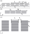
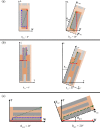
Bone tissue at nanoscale is a composite mainly made of apatite crystals, collagen molecules and water. This work is aimed to study the diffusion within bone nanostructure through Monte-Carlo simulations. To this purpose, an idealized geometric model of the apatite-collagen structure was developed. Gaussian probability distribution functions were employed to design the orientation of the apatite crystals with respect to the axes (length L, width W and thickness T) of a plate-like trabecula. We performed numerical simulations considering the influence of the mineral arrangement on the effective diffusion coefficient of water. To represent the hindrance of the impermeable apatite crystals on the water diffusion process, the effective diffusion coefficient was scaled with the tortuosity, the constrictivity and the porosity factors of the structure. The diffusion phenomenon was investigated in the three main directions of the single trabecula and the introduction of apatite preferential orientation allowed the creation of an anisotropic medium. Thus, different diffusivities values were observed along the axes of the single trabecula. We found good agreement with previous experimental results computed by means of a genetic algorithm.
Conflict of interest statement
Figures


Similar articles
-
A 3D Model of the Effect of Tortuosity and Constrictivity on the Diffusion in Mineralized Collagen Fibril.Sci Rep. 2019 Feb 25;9(1):2658. doi: 10.1038/s41598-019-39297-w. Sci Rep. 2019. PMID: 30804401 Free PMC article.
-
Experimental study of diffusion coefficients of water through the collagen: apatite porosity in human trabecular bone tissue.Biomed Res Int. 2014;2014:796519. doi: 10.1155/2014/796519. Epub 2014 May 21. Biomed Res Int. 2014. PMID: 24967405 Free PMC article.
-
Trabecular health of vertebrae based on anisotropy in trabecular architecture and collagen/apatite micro-arrangement after implantation of intervertebral fusion cages in the sheep spine.Bone. 2018 Mar;108:25-33. doi: 10.1016/j.bone.2017.12.012. Epub 2017 Dec 11. Bone. 2018. PMID: 29241826
-
[Anisotropic crystallization of biominerals].Clin Calcium. 2014 Feb;24(2):193-202. Clin Calcium. 2014. PMID: 24473352 Review. Japanese.
-
Bone strength and residual compressive stress in apatite crystals.J Struct Biol. 2024 Dec;216(4):108141. doi: 10.1016/j.jsb.2024.108141. Epub 2024 Oct 22. J Struct Biol. 2024. PMID: 39442775 Review.
Cited by
-
Mathematical modeling of vancomycin release from Poly-L-Lactic Acid-Coated implants.PLoS One. 2024 Nov 1;19(11):e0311521. doi: 10.1371/journal.pone.0311521. eCollection 2024. PLoS One. 2024. PMID: 39485770 Free PMC article.
-
Harnessing Biofabrication Strategies to Re-Surface Osteochondral Defects: Repair, Enhance, and Regenerate.Biomimetics (Basel). 2023 Jun 15;8(2):260. doi: 10.3390/biomimetics8020260. Biomimetics (Basel). 2023. PMID: 37366855 Free PMC article. Review.
-
Percolation networks inside 3D model of the mineralized collagen fibril.Sci Rep. 2021 May 31;11(1):11398. doi: 10.1038/s41598-021-90916-x. Sci Rep. 2021. PMID: 34059767 Free PMC article.
-
Uncovering a high-performance bio-mimetic cellular structure from trabecular bone.Sci Rep. 2020 Aug 28;10(1):14247. doi: 10.1038/s41598-020-70536-7. Sci Rep. 2020. PMID: 32859928 Free PMC article.
-
3D Tortuosity and Diffusion Characterization in the Human Mineralized Collagen Fibril Using a Random Walk Model.Bioengineering (Basel). 2023 May 7;10(5):558. doi: 10.3390/bioengineering10050558. Bioengineering (Basel). 2023. PMID: 37237628 Free PMC article.
References
-
- Marinozzi F, Marinozzi A, Bini F, Zuppante F, Pecci R, Bedini R. Variability of morphometric parameters of human trabecular tissue from coxo-arthritis and osteoporotic samples. Ann I Super Sanità 2012; 48: 19–25. - PubMed
-
- Marinozzi F, Bini F, Marinozzi A, Zuppante F, De Paolis A, Pecci R et al. Technique for bone volume measurement from human femur head samples by classification of micro-CT image histograms. Ann I Super Sanità 2013; 49: 300–305. - PubMed
-
- Fratzl P, Gupta HS, Paschalis EP, Roschger P. Structure and mechanical quality of the collagen-mineral nano-composite in bone. J Mater Chem 2004; 14: 2115–2123.
-
- Weiner S, Wagner HD. The material bone: Structure-mechanical function relations. Annu Rev Mater Sci 1998; 28: 271–298.
-
- Rho JY, Kuhn-Spearing L, Zioupos P. Mechanical properties and the hierarchical structure of bone. Med Eng Phys 1998; 20: 92–102. - PubMed
MeSH terms
Substances
LinkOut - more resources
Full Text Sources
Other Literature Sources

