. 2017 Dec 8;12(12):e0189211.
doi: 10.1371/journal.pone.0189211. eCollection 2017.

Hexahydrocurcumin protects against cerebral ischemia/reperfusion injury, attenuates inflammation, and improves antioxidant defenses in a rat stroke model

Affiliations

Hexahydrocurcumin protects against cerebral ischemia/reperfusion injury, attenuates inflammation, and improves antioxidant defenses in a rat stroke model

Piyawadee Wicha et al. PLoS One. .

Abstract

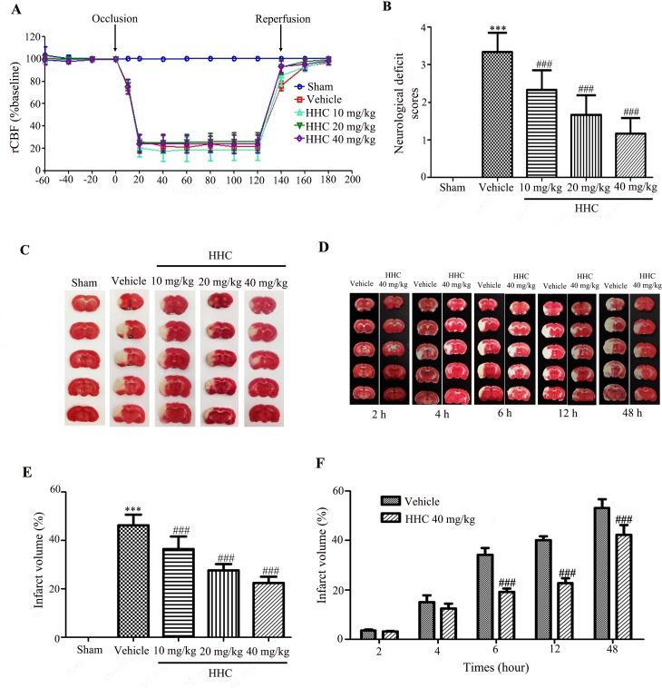
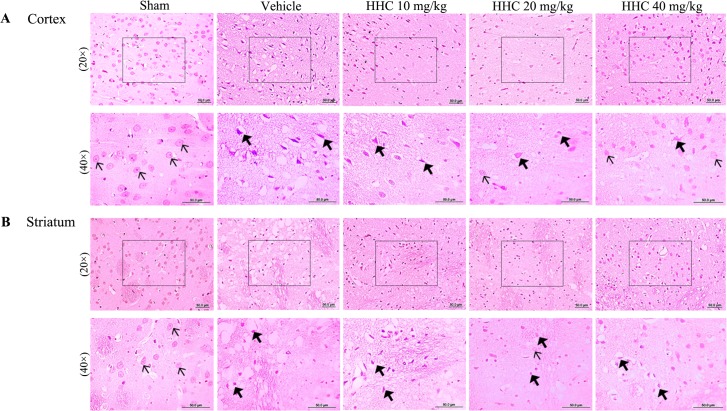
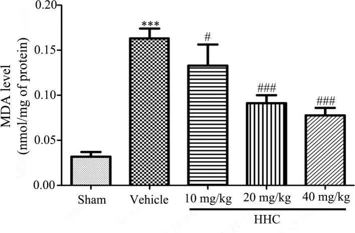
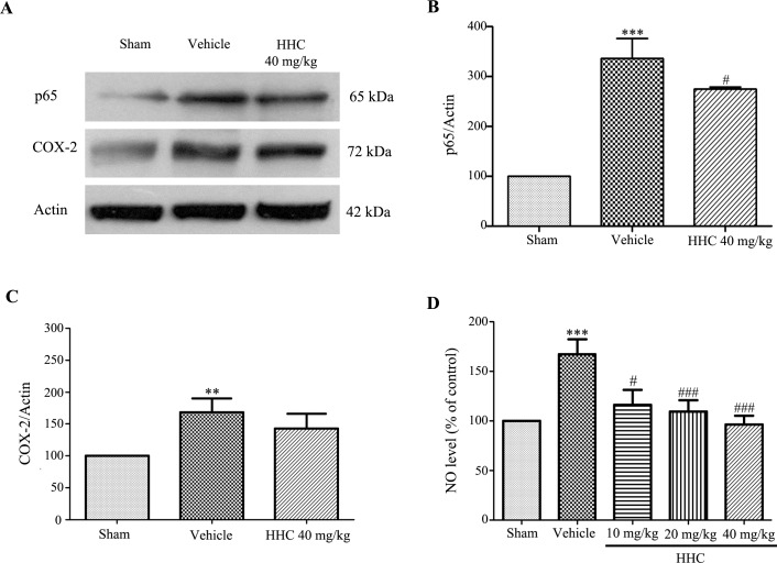
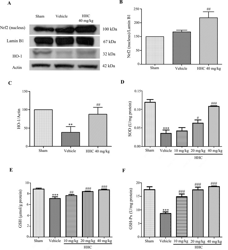
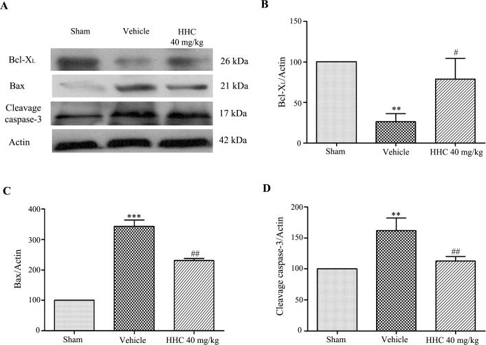
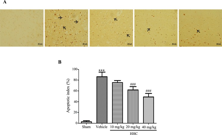
The purpose of the present experiment was to investigate whether hexahydrocurcumin (HHC) attenuates brain damage and improves functional outcome via the activation of antioxidative activities, anti-inflammation, and anti-apoptosis following cerebral ischemia/reperfusion (I/R). In this study, rats with cerebral I/R injury were induced by a transient middle cerebral artery occlusion (MCAO) for 2 h, followed by reperfusion. The male Wistar rats were randomly divided into five groups, including the sham-operated, vehicle-treated, 10 mg/kg HHC-treated, 20 mg/kg HHC-treated, and 40 mg/kg HHC-treated I/R groups. The animals were immediately injected with HHC by an intraperitoneal administration at the onset of cerebral reperfusion. After 24 h of reperfusion, the rats were tested for neurological deficits, and the pathology of the brain was studied by 2,3,5-triphenyltetrazolium chloride (TTC) staining, hematoxylin and eosin (H&E) staining, and terminal deoxynucleotidyltransferase UTP nick end labeling (TUNEL) staining. In addition, the brain tissues were prepared for protein extraction for Western blot analysis, a malondialdehyde (MDA) assay, a nitric oxide (NO) assay, a superoxide dismutase (SOD) assay, a glutathione (GSH) assay, and a glutathione peroxidase (GSH-Px) assay. The data revealed that the neurological deficit scores and the infarct volume were significantly reduced in the HHC-treated rats at all doses compared to the vehicle group. Treatment with HHC significantly attenuated oxidative stress and inflammation, with a decreased level of MDA and NO and a decreased expression of NF-κB (p65) and cyclooxygenase-2 (COX-2) in the I/R rats. HHC also evidently increased Nrf2 (nucleus) protein expression, heme oxygenase-1 (HO-1) protein expression, the antioxidative enzymes, and the superoxide dismutase (SOD) activity. Moreover, the HHC treatment also significantly decreased apoptosis, with a decrease in Bax and cleaved caspase-3 and an increase in Bcl-XL, which was in accordance with a decrease in the apoptotic neuronal cells. Therefore, the HHC treatment protects the brain from cerebral I/R injury by diminishing oxidative stress, inflammation, and apoptosis. The antioxidant properties of HHC may play an important role in improving functional outcomes and may offer significant neuroprotection against I/R damage.


Conflict of interest statement

Competing Interests: The authors have declared that no competing interests exist.

Figures

Fig 1
Fig 1. Effects of HHC on brain infarction and neurological outcomes after cerebral I/R in rats.
HHC was administered upon initiation of the reperfusion. (A) The regional cerebral blood flow (rCBF) observed during pre-occlusion, occlusion, and reperfusion. (B) The neurological deficit scores after cerebral I/R. (C) The TTC staining of series coronal brain section (2 mm thick) after 24-h reperfusion. The infarct region is in white color, whereas the non-ischemic region appears in red color. (D) The TTC staining of rat treatment with 40 mg/kg HHC and vehicle group at 2 h, 4 h, 6 h, 12 h and 48 h after reperfusion. (E) The percentage of infarct volume after I/R in different groups. (F) The percentage of infarct volume after I/R in various reperfusion time. All the data are presented as mean±SD. *** P < 0.001 versus sham group. ### P < 0.001 versus vehicle group.
Fig 2
Fig 2. The representative histopathological changes of the brain at 24 h after focal cerebral I/R in stroke rats.
The brain tissues were stained with H&E and visualized with a light microscope (20× and 40×). In the sham group, the structure of most of the neurons in the cortex and the striatum were clear (thin arrow). The neurons of the I/R group were found to present shrinkage, chromosome condensation, and nuclear pyknosis, and showed increased intercellular space (thick arrow). The number of intact neurons in the rats that were treated with HHC was found to have increased.
Fig 3
Fig 3. HHC reduces markers of lipid peroxidation in the ischemic tissue.
The effects of HHC on the MDA level in different groups. The data are presented as mean±SD (n = 6). *** P < 0.001versus sham group. # P < 0.05 versus vehicle group, and ### P < 0.001 versus vehicle group.
Fig 4
Fig 4. The effect of HHC on inflammation in cerebral I/R rats.
(A) Representative of the NF-κB (p65) and the COX-2 protein expressions, as examined by western blot analysis. (B) The quantitative results of the expressions of the NF-κB (p65) proteins in each of the groups. (C) The quantitative results of the expressions of the COX-2 proteins in each of the groups. (D) The effect of HHC on the NO level in stroke rats. The data are expressed as mean±SD. ** P < 0.01 versus sham group, and *** P < 0.001 versus sham group. # P < 0.05 versus vehicle group, and ### P < 0.001 versus vehicle group.
Fig 5
Fig 5. The effect of HHC on the Nrf2 signaling pathway in cerebral I/R injury rats.
(A) Representative of the Nrf2 (nucleus) and the HO-1 protein expressions, as examined by western blotting. (B) The quantitative results of the expressions of the Nrf2 proteins in each of the groups. (C) The quantitative results of the expressions of the HO-1 proteins in each of the groups. (D) The effect of HHC on SOD activity in cerebral I/R injury rats. (E) The effect of HHC on GSH in cerebral I/R injury rats. (F) The effect of HHC on GSH-Px in cerebral I/R injury rats. The data are presented as mean±SD. ** P < 0.01 versus sham group, and *** P < 0.001 versus sham group. # P < 0.05 versus vehicle group, ## P < 0.01 versus vehicle group, and ### P < 0.001 versus vehicle group.
Fig 6
Fig 6. The effect of HHC on the apoptosis pathway in stroke rats.
(A) Representative of the Bax, the Bcl-XL, and the cleaved caspase-3 protein expressions in the penumbra cortex, as determined by western blot analysis. (B) The quantitative results of the expressions of the Bcl-XL proteins in each of the groups. (C) The quantitative results of the expressions of the Bax protein in each of the groups. (D) The quantitative results of the expressions of the cleaved caspase-3 protein in each of the groups. The data are presented as mean±SD. ** P < 0.01 versus sham group, and *** P < 0.001 versus sham group. # P < 0.05 versus vehicle group and ## P < 0.01 versus vehicle group.
Fig 7
Fig 7. The effect of HHC on I/R-induced neuronal apoptosis.
(A) The apoptotic cells examined by TUNEL staining and which were visualized with a light microscope (20×). The arrows show TUNEL-positive cells. (B) The quantitative analysis of TUNEL-positive cells in each of the groups. The data are presented as mean±SD. ***P < 0.001 versus sham group. ### P < 0.001 versus vehicle group.

References

    1. Donnan GA, Fisher M, Macleod M, Davis SM. Stroke. Lancet. 2008; 371: 1612–1623. doi: 10.1016/S0140-6736(08)60694-7 - DOI - PubMed
    1. Hicks A, Jolkkonen J. Challenges and possibilities of intravascular cell therapy in stroke. Acta Neurobiol Exp (Wars). 2009; 69: 1–11. - PubMed
    1. Rahme R, Curry R, Kleindorfer D, Khoury JC, Ringer AJ, Kissela BM, et al. How often are patients with ischemic stroke eligible for decompressive hemicraniectomy?. Stroke. 2012; 43: 550–552. doi: 10.1161/STROKEAHA.111.635185 - DOI - PMC - PubMed
    1. Fujimura M, Gasche Y, Morita-Fujimura Y, Massengale J, Kawase M, Chan PH. Early appearance of activated matrix metalloproteinase-9 and blood-brain barrier disruption in mice after focal cerebral ischemia and reperfusion. Brain Res. 1999; 84: 92–100. PMID: 10526099 - PubMed
    1. Fukuda AM, Badaut J. Aquaporin 4: a player in cerebral edema and neuroinflammation. J Neuroinflammation. 2012; 279: 2094–2099. doi: 10.1186/1742-2094-9-279 PMID: 23270503 - DOI - PMC - PubMed