. 2017 Dec 7;68(5):955-969.e10.
doi: 10.1016/j.molcel.2017.11.013.

hnRNPK Recruits PCGF3/5-PRC1 to the Xist RNA B-Repeat to Establish Polycomb-Mediated Chromosomal Silencing

Affiliations

hnRNPK Recruits PCGF3/5-PRC1 to the Xist RNA B-Repeat to Establish Polycomb-Mediated Chromosomal Silencing

Greta Pintacuda et al. Mol Cell. .

Abstract

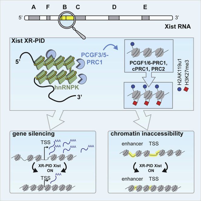
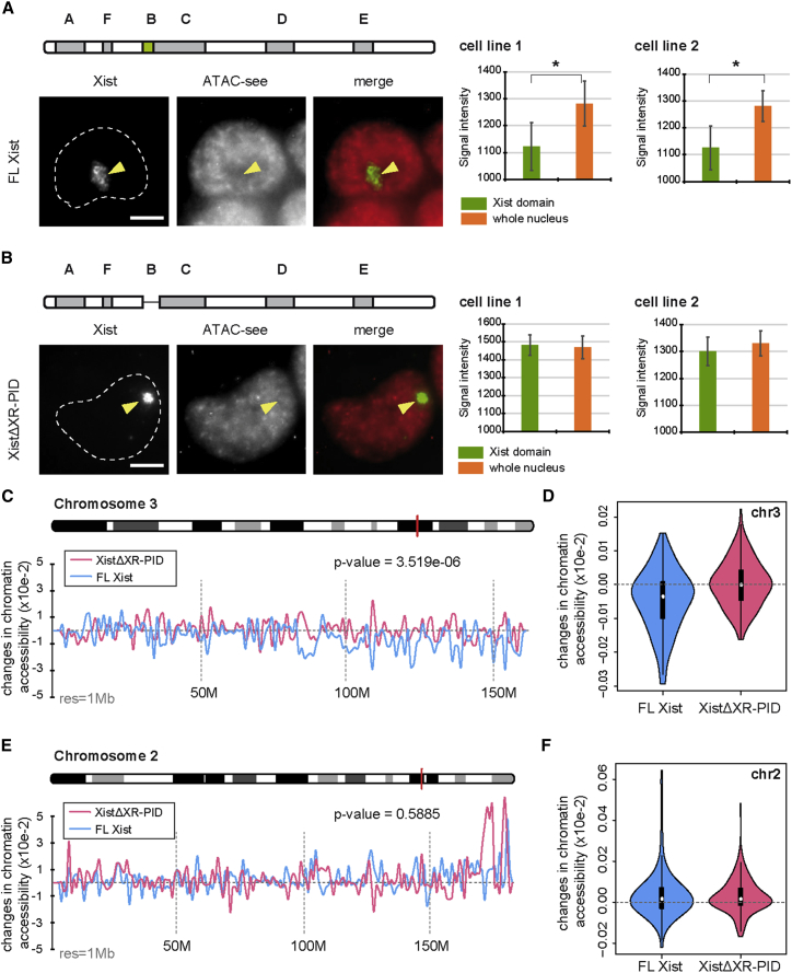
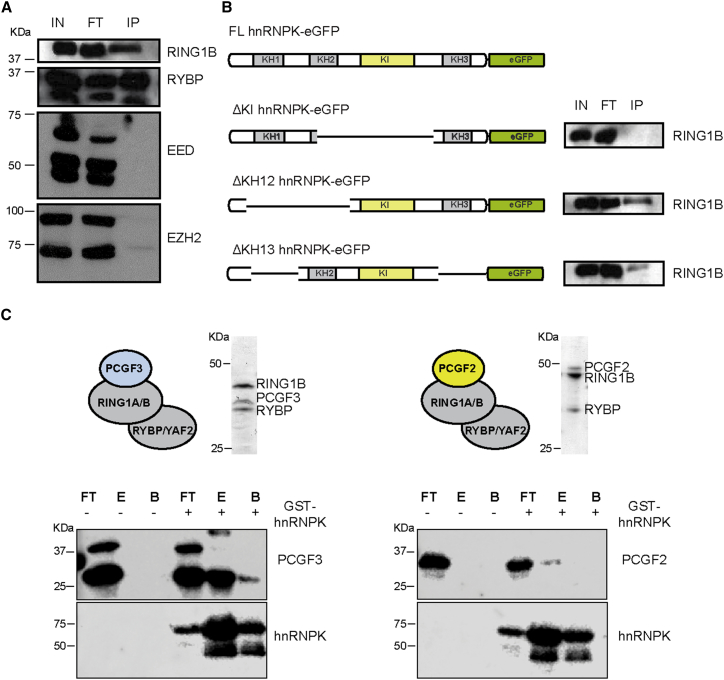
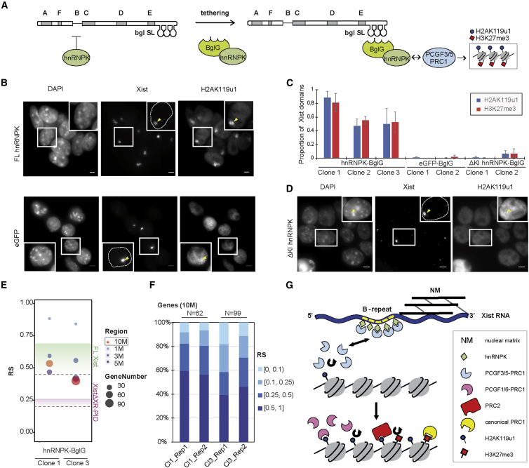
The Polycomb-repressive complexes PRC1 and PRC2 play a key role in chromosome silencing induced by the non-coding RNA Xist. Polycomb recruitment is initiated by the PCGF3/5-PRC1 complex, which catalyzes chromosome-wide H2A lysine 119 ubiquitylation, signaling recruitment of other PRC1 complexes, and PRC2. However, the molecular mechanism for PCGF3/5-PRC1 recruitment by Xist RNA is not understood. Here we define the Xist RNA Polycomb Interaction Domain (XR-PID), a 600 nt sequence encompassing the Xist B-repeat element. Deletion of XR-PID abolishes Xist-dependent Polycomb recruitment, in turn abrogating Xist-mediated gene silencing and reversing Xist-induced chromatin inaccessibility. We identify the RNA-binding protein hnRNPK as the principal XR-PID binding factor required to recruit PCGF3/5-PRC1. Accordingly, synthetically tethering hnRNPK to Xist RNA lacking XR-PID is sufficient for Xist-dependent Polycomb recruitment. Our findings define a key pathway for Polycomb recruitment by Xist RNA, providing important insights into mechanisms of chromatin modification by non-coding RNA.


Figures

None
Graphical abstract
Figure 1
Figure 1
A 0.6 kb Element, XR-PID, Mediates Polycomb Recruitment by Xist RNA (A) Schematic illustrating the XistΔXEv deletion (encompassing XR-PID) relative to FL-Xist. The location of Xist tandem repeats A–F is indicated. The XistΔXN transgene described previously is shown for comparison. (B) Examples of immunoFISH analysis illustrating presence or absence of H2AK119u1 foci (arrowheads), following 24 hr induction of Xist transgenes. Images are stacks of six consecutive Z sections with insets showing a single section. Nuclei outlines determined from DAPI stain are indicated with dashed line in inset. Scale bar indicates 5 μm. (C) Quantitative analysis of a single FL-Xist and XistΔXEv cell line based on scoring Xist RNA domains for presence or absence of H2AK119u1 or H3K27me3. Error bars represent SD for at least three biological replicates with a sample of n > 100 for each replicate. (D) As in (A). Green shading highlights B-repeat element. (E) As in (B). (F) As in (C). ∗∗p < 0.001 relative to FL-Xist (2 tailed Student’s t test).
Figure 2
Figure 2
The XR-PID Element Contributes to Transcriptional Silencing Mediated by Xist RNA (A) RS distribution across all autosomes in two biological replicates, for two cell lines expressing FL-Xist (left) versus XistΔXR-PID (right), following a 72 hr induction of the transgenes. (B) RS distribution in continuous 10 Mb windows across chromosome 3 for FL-Xist (left) and chromosome 2 for XistΔXR-PID (right). Purple dots represent the RS for each gene. Blue boxes indicate maximum RS window. (C) UCSC genome browser tracks showing the Cnn3 locus (left) and Dstn locus (right) after induction of FL-Xist or XistΔXR-PID, respectively. Gray tracks represent no-dox control. Red and blue are two biological replicates with 72 hr dox treatment. The active allele (129S1) and inactive allele (Cast) are indicated. Chromosomal location of Cnn3 and Dstn is indicated with a red bar on the chromosome ideogram above each example. (D) Comparison of RS in windows centered on maximal RS for cells harboring FL-Xist and Xist transgenes as indicated. The color and area of circles indicate the size and gene number within the region, respectively. (E) RS comparison (% of genes) in the 10 Mb of maximum silencing windows depicted in (B). RS range is as indicated.
Figure 3
Figure 3
XR-PID-Mediated Silencing Analyzed using EvXist, a Truncated Xist Transgene (A) Schematic for site-specific integration of a single copy of EvXist or EvXistΔXR-PID into the Col1a1 locus (in red) on chromosome (chr) 11 (top). Heatmap shows the RS in each chromosome for both replicates (rep), using EvXist or EvXistΔXR-PID (bottom). (B) RS distribution in continuous 10 Mb windows across chromosome 11, following 72 hr induction of single-copy EvXist (top) or EvXistΔXR-PID (bottom) transgenes. Purple dot represents the RS for each gene. The position of the Col1a1 locus is indicated. (C) RS comparison between EvXist and EvXistΔXR-PID in windows centered on the region of maximum silencing. The color and area of dots indicate the region size and gene number, respectively. (D) RS comparison (% of genes) between EvXist and EvXistΔXR-PID cell lines in the 10 Mb of maximum silencing windows depicted in (B). The RS was grouped into four categories, indicated by the graded color. (E) Scatterplot showing the differences of the calibrated (c) RS for 454 repressed genes in EvXist and EvXistΔXR-PID. The axis represents log2-transformed cRS. (F) Heatmap illustrating RS of each gene ranked by genomic coordinates on chromosome 11 for both biological replicates of EvXist and EvXistΔXR-PID cells (left). The Syngr2-Tk1 locus, indicated by red lines, is shown in the upper chromosome ideogram. RS is indicated, and q value for both Syngr2 and Tk1 is 0 in EvXist cells and 0.036 and 0.611, respectively, in EvXistΔXR-PID cells. Data for no dox (untreated) and for two replicates (rep) of 72 hr induction (red and blue) are shown. Active (129S1) and inactive (Cast) alleles are labeled (right).
Figure 4
Figure 4
XR-PID Is Required for Reduced Chromatin Accessibility over Xist Domains (A and B) Individual examples illustrating Xist RNA-FISH combined with ATAC-see. The nuclei (DAPI staining) are outlined with a dotted line with Xist domains indicated (arrowhead). Scale bar indicates 5 μm. Fluorescence intensity of the ATAC-see signal overlapping Xist and total nuclear signal were measured for two independent cell lines for FL-Xist (A) and XistΔXR-PID transgenes. Bar graphs represent average values for three biological replicates (n > 20) for each independent cell line. Error bars represent SD. p < 0.05 (paired Student t test). (C) Chromatin accessibility changes upon FL-Xist induction on chromosome 3. The red bar on the ideogram indicates maximum silencing regions. The chr3-wide accessibility changes from XistΔXR-PID expressing cells, in which the Xist transgene is on chr2, serves as a control. The p value was calculated using Mann-Whitney test. (D) Chromatin accessibility changes in the presence (blue) or absence (red) of FL-Xist expression. (E) Chromatin accessibility changes upon XistΔXR-PID induction on chromosome 2. Red bar on the ideogram indicates maximum silencing regions. The chr2-wide accessibility changes from FL-Xist transgene cells (chr3) provide a control. The p value was calculated using Mann-Whitney test. (F) As in (D) for XistΔXR-PID.
Figure 5
Figure 5
hnRNPK Binds Xist B-Repeat (A and B) Schematic (A) of the proteomic screening strategy using different in vitro transcribed Xist constructs, indicated in (B). Proteins in nuclear extract (gray shapes) that bind in vitro-transcribed RNA with incorporated biotin (filled lollipop) were captured by UV cross-linking and then purified using streptavidin beads (large filled circle) prior to analysis by MS. (C) Overlap with Xist-interacting partners identified in independent biochemical (Chu et al., 2015) and genetic (Moindrot et al., 2015) screens. (D and E) Scatterplots representing abundance of factors interacting preferentially with the A-repeat (D) or the B-repeat (E) elements of Xist by quantitative MS. Q value was calculated across two biological replicates. (F) Western blot shows levels of hnRNPK before and after 24, 48, and 72 hr siRNA treatment. Histone H3 was used as a loading control. Bar chart quantifies immunoFISH analysis of H2AK119u1 and Xist RNA co-localization in cells treated with hnRNPK or scrambled (control) siRNA. Data are from two independent siRNA transfections, each with three biological replicates (n > 50). Error bars represent SD across all biological replicates. (G) UCSC track displaying hnRNPK iCLIP data (Cirillo et al., 2016) aligned to Xist RNA sequence. The correspondence with the XR-PID and specifically with the B-repeat core motif is highlighted in light blue. The most prominent peak of hnRNPK binding is enlarged below (red) and aligned to the genomic sequence of the XR-PID across several mammalian species. The consensus B-repeat core sequence spanning the region is also represented.
Figure 6
Figure 6
hnRNPK Interacts Directly with PCGF3/5-PRC1 (A) Western blots illustrating eGFP pull-down of PRC1 and PRC2 proteins (as indicated) in cells expressing hnRNPK-eGFP. Loaded were 0.1% of the input (IN), 0.1% of the flowthrough (FT), and 10% of the pull-down (IP). (B) RING1B pull-down repeated using cell lines expressing hnRNPK-eGFP deletion variants as indicated in schematic. Loading is as in (A). (C) Western blots illustrating pull-down of recombinant PCGF3-PRC1, but not PCGF2-PRC1, using recombinant GST-hnRNPK. A Coomassie brilliant blue-stained gel of the complexes used in the assay is shown above. Loaded were 0.1% of flowthrough (FT), 20% of eluate (E), and 33% of bead bound (B).
Figure 7
Figure 7
Tethering hnRNPK Is Sufficient for Xist-Dependent Polycomb Recruitment (A) Schematic illustrating the hnRNPK-tethering experiment. (B) Examples of ImmunoFISH detection of Xist RNA and H2AK119u1 using BglG-hnRNPK or BglG-eGFP. Wide-field images represent stacks of 10 consecutive Z sections. Arrows indicate Xist domains. Insets showing enlarged individual cells are single z sections. Scale bar indicates 5 μm. (C) Bar graphs illustrate quantification for H2AK119u1 and H3K27me3 domains overlapping Xist in independent cell lines (clones) of each named construct. Error bars represent SD across at least three biological replicates (n > 100). (D) Example as in (B) for BglG-ΔKI hnRNPK. (E) RS comparison between two independent BglG-hnRNPK cell lines in windows centered on the region of maximum silencing. The color and area of dots indicate the region size and gene number, respectively. Green and pink gradients represent the range of RS previously measured for FL and XistΔXR-PID lines, respectively. (F) RS comparison between two independent BglG-hnRNPK cell lines in the 10 Mb of maximum silencing windows. The RS was grouped into four categories, indicated by the graded color. (G) Model illustrating molecular pathway for Polycomb recruitment by Xist RNA.

Similar articles

Cited by

References

    1. Almeida M., Pintacuda G., Masui O., Koseki Y., Gdula M., Cerase A., Brown D., Mould A., Innocent C., Nakayama M. PCGF3/5-PRC1 initiates Polycomb recruitment in X chromosome inactivation. Science. 2017;356:1081–1084. - PMC - PubMed
    1. Anders S., Pyl P.T., Huber W. HTSeq—a Python framework to work with high-throughput sequencing data. Bioinformatics. 2015;31:166–169. - PMC - PubMed
    1. Arnold P., Schöler A., Pachkov M., Balwierz P.J., Jørgensen H., Stadler M.B., van Nimwegen E., Schübeler D. Modeling of epigenome dynamics identifies transcription factors that mediate Polycomb targeting. Genome Res. 2013;23:60–73. - PMC - PubMed
    1. Arrigoni R., Alam S.L., Wamstad J.A., Bardwell V.J., Sundquist W.I., Schreiber-Agus N. The Polycomb-associated protein Rybp is a ubiquitin binding protein. FEBS Lett. 2006;580:6233–6241. - PubMed
    1. Beard C., Hochedlinger K., Plath K., Wutz A., Jaenisch R. Efficient method to generate single-copy transgenic mice by site-specific integration in embryonic stem cells. Genesis. 2006;44:23–28. - PubMed

MeSH terms