. 2017 Dec 7;101(6):1021-1033.
doi: 10.1016/j.ajhg.2017.11.006.

ACTB Loss-of-Function Mutations Result in a Pleiotropic Developmental Disorder

Affiliations

ACTB Loss-of-Function Mutations Result in a Pleiotropic Developmental Disorder

Sara Cuvertino et al. Am J Hum Genet. .

Abstract

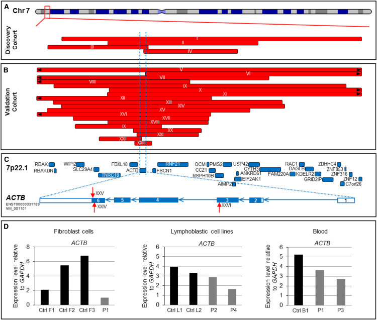
ACTB encodes β-actin, an abundant cytoskeletal housekeeping protein. In humans, postulated gain-of-function missense mutations cause Baraitser-Winter syndrome (BRWS), characterized by intellectual disability, cortical malformations, coloboma, sensorineural deafness, and typical facial features. To date, the consequences of loss-of-function ACTB mutations have not been proven conclusively. We describe heterozygous ACTB deletions and nonsense and frameshift mutations in 33 individuals with developmental delay, apparent intellectual disability, increased frequency of internal organ malformations (including those of the heart and the renal tract), growth retardation, and a recognizable facial gestalt (interrupted wavy eyebrows, dense eyelashes, wide nose, wide mouth, and a prominent chin) that is distinct from characteristics of individuals with BRWS. Strikingly, this spectrum overlaps with that of several chromatin-remodeling developmental disorders. In wild-type mouse embryos, β-actin expression was prominent in the kidney, heart, and brain. ACTB mRNA expression levels in lymphoblastic lines and fibroblasts derived from affected individuals were decreased in comparison to those in control cells. Fibroblasts derived from an affected individual and ACTB siRNA knockdown in wild-type fibroblasts showed altered cell shape and migration, consistent with known roles of cytoplasmic β-actin. We also demonstrate that ACTB haploinsufficiency leads to reduced cell proliferation, altered expression of cell-cycle genes, and decreased amounts of nuclear, but not cytoplasmic, β-actin. In conclusion, we show that heterozygous loss-of-function ACTB mutations cause a distinct pleiotropic malformation syndrome with intellectual disability. Our biological studies suggest that a critically reduced amount of this protein alters cell shape, migration, proliferation, and gene expression to the detriment of brain, heart, and kidney development.

Keywords: ACTB; chromatin; developmental disorder; malformations; β-actin.


Figures

Figure 1
Figure 1
ACTB Loss-of-Function Mutations Result in Reduced Expression (A) Discovery cohort—a representation of chromosome 7 with four small 7p22.1 deletions identified in the discovery cohort. The red bars represent the extent of genomic deletions, and the associated family number is linked to Table 1. (B) Validation cohort—the 19 small 7p22.1 deletions identified in the validation cohort. The red bars represent the extent of genomic deletions, and the associated family number is linked to Table 1. (C) Intragenic ACTB mutations—the location of the known protein-coding genes on 7p22.1 are shown in blue boxes. ACTB is highlighted by two flanking dashed blue lines. The lower panel shows the exon structure of ACTB. The boxes represent the ACTB exons, and the blue arrows denote the introns. Note that ACTB is transcribed from the reverse strand. The filled (blue) and unfilled sections of the exons denote translated and untranslated regions of the gene, respectively. Location of mutations in the three affected individuals is shown with red arrows—one with a small frameshift deletion (XXIV: p.Ser368LeufsTer13), one with a nonsense mutation (XXV: p.Lys373Ter) and one with de novo frameshift mutation (XXVI: p.Leu110ArgfsTer10). The transcript ID is NM_001101.3. (D) ACTB loss-of-function mutations result in reduced gene expression—quantitative real-time polymerase chain reaction (qRT-PCR) analysis of ACTB transcript levels relative to GAPDH in fibroblasts, LCL and blood samples (sample P1 is from IVa, P2 is from XI, P3 is from II, and P4 is from XXII). Cells were collected by centrifugation, and total RNAs were extracted with the RNeasy Mini kit (QIAGEN) according to the manufacturer’s protocol. The qRT-PCR reactions were performed on a Bio-Rad CFX394 Real-Time system (Bio-Rad) with Power SYBR Green PCR Master mix (Applied Biosystems). The expression of each target gene was evaluated via a relative quantification approach (-ΔCT method), and GAPDH was used as the internal reference for human genes.
Figure 2
Figure 2
ACTB Loss-of-Function Mutations Result in a Recognizable Phenotype (A) Facial gestalt and physical anomalies with ACTB loss-of-function mutations—the facial features of a number of individuals who had 7p22.1 deletions or point mutations (marked with ) in ACTB were remarkably similar to each other; they had wavy interrupted eyebrows, dense eyelashes, a wide nose, a wide mouth, and a prominent chin. Several affected individuals in this cohort demonstrated overlapping toes, small nails, and spinal anomalies such as sacral dimples. (B) Immunohistochemistry of embryonic day 14 mice. The top row shows β-actin immunostaining (brown) in neurons in the brain (neocortex), epithelia of kidney tubules (S-shaped body), and the heart (endocardium and cardiac outflow). The bottom row shows sections with primary antibody omitted so that the specificity of the above signals is shown. Whole embryos at day 14 (E14) were fixed in 4% paraformaldehyde and embedded in paraffin. Sections of 5 μm were cut and mounted on polylysine-coated glass slides. Endogenous peroxidase was quenched by incubation with hydrogen peroxide (0.3% solution in PBS). Embryos were heated at 95°C for 5 minutes in sodium citrate (pH 6) for antigen exposure. Rabbit anti-β-actin (1:250, Abcam ab8227) was applied to tissue sections over night at 4°C. Goat anti-rabbit (1:200) was applied for 1 hr at room temperature and revealed with the ABC Elite kit (Vector) followed by DAB staining (Vector) and hematoxylin counter-stain (scale bar: 50 μm).
Figure 3
Figure 3
ACTB Loss-of-Function Mutations Induce Abnormalities of Cellular Morphology and Reduced Migration (A) Immunoblots of cytoplasmic β-actin. No consistent differences were detected in the cytoplasmic β-actin amounts in the fibroblasts and LCLs of affected individuals versus controls (sample P1 is from IVa, P2 is from XI, P3 is from II, and P4 is from XXII). Immunoblotting for β-actin was performed on the cytoplasmic protein fraction, and GAPDH was used as a loading control. Protein samples were isolated using NE-PER nuclear and cytoplasmic extraction reagents (ThermoScientific). 8–10 mg of protein extracts were loaded into the polyacrylamide gel Bolt 10% Bis-Tris Plus Gels (Invitrogen). The membranes were incubated with specific anti-beta actin (ab8227, Abcam) and anti-GAPDH (5174S, Cell Signaling) overnight at 4°C. After washes, the membranes were incubated with a secondary fluorescently labeled goat anti-rabbit antibody (IRDye 800CW Li-Cor), and signal was developed with an Odyssey CLX imaging machine. (B) Fibroblast morphology. ACTB-deficient cells were found to be significantly more circular than non-deficient cells. Phalloidin and DAPI immunostaining was performed in wild-type fibroblasts transfected with 30 nM control siRNA (ON-TARGETplus non-targeting pool, Fisher) and ACTB siRNA (SMARTpool ON TARGET plus ACTB siRNA, Dharmacon) and affected-individual fibroblasts transfected with control siRNA. Cells were fixed with 4% paraformaldehyde for 15 minutes at room temperature. After blocking solution was washed out, antibody Texas Red-X Phalloidin (T7471, Life Technologies) was applied for 1 hr at room temperature in the dark. Samples were stained with DAPI (4083S, Cell Signaling Technology) for 5 min. Representative pictures show marked difference in the morphology of β-actin-deficient cells (scale bar: 50 μm). Enlargement of cells is shown in the lower panels. Dashed lines show the outline of the cell boundary. The left bar chart shows that there was no significant difference in the area of each of the cell groups (in μm2) as calculated with ImageJ software. The right bar chart shows the increased circularity of the ACTB-deficient fibroblasts as calculated with ImageJ software (n = 4; ∗∗p < 0.01). Values of 1 and 0 stand for a perfect circle and a line, respectively. Error bars indicate mean ± 1 SD. (C) Fibroblast migration. ACTB-deficient cells had impaired migration. A migration assay was performed in wild-type fibroblasts transfected with control siRNA and ACTB siRNA and affected-individual fibroblasts transfected with control siRNA. 96 hr after transfection, a wound was generated in the confluent monolayer of fibroblasts via a p200 pipet tip. Cells were washed with phosphate-buffered saline so that any debris created by the wound would be removed. The first image of the wound was taken with a phase-contrast microscope, and marking the plate under the capture image field created a reference point. Cells were incubated at 37°C in a humidified 5% CO2 incubator for 2 days. After the incubation time, a second image was taken. For quantifying the migration of cells, the cells that crossed into the wound area were counted. Representative pictures show reduced migration in β-actin-deficient cells (scale bar: 100 μm). The bar chart shows that the numbers of cells in the central wound area are significantly lower in the β-actin-deficient cells. Data are shown as the mean of absolute cell numbers at 144 hours from two wells of two independent experiments (n = 4; p < 0.05, ∗∗p < 0.01). Error bars indicate mean ± 1 SD.
Figure 4
Figure 4
ACTB Loss-of-Function Mutations Lead to Reduced Expression of β-actin in the Nucleus, Reduced Cell Proliferation, and Dysregulation of Cell-Cycle Genes (A) Immunoblots of nuclear β-actin. Nuclear β-actin amounts in fibroblasts and LCLs from affected individuals were consistently lower than those of their respective controls (sample P1 is from IVa, P2 is from XI, P3 is from II, and P4 is from XXII). Immunoblotting for β-actin (ab8227, Abcam) was performed on the nuclear protein fraction with histone 3 (ab1791, Abcam) as a loading control. Visual inspection of the membranes and their intensity quantification revealed a consistent trend of decreased amounts of nuclear β-actin in fibroblasts and LCLs from affected individuals versus controls (sample P1 is from IVa, P2 is from XI, P3 is from II, and P4 is from XXII). (B) Fibroblast and LCL proliferation. Proliferation in cells derived from affected individuals was found to be significantly reduced. Quantification of the number of fibroblasts and LCLs was performed in a 12-well plate. Cells were plated in the presence of their growth medium and counted with a hemocytometer every 3 days for 9 days. Fibroblasts and LCLs from affected individuals proliferated significantly slowly in comparison with the control cells. Data are shown as the mean of absolute cell number from three wells of three independent experiments (n = 3; p < 0.05, ∗∗p < 0.01). Error bars indicate mean ± 1 SD. (C) Expression of cell-cycle genes in LCLs. Cell-cycle genes were dysregulated in LCLs from affected individuals. The abundance of selected mRNAs in LCLs derived from individuals XI (P2) and XXII (P4) is shown relative to mean amounts in LCLs in two control individuals; measurements are in FPKM (fragments per kilo base of transcript per million mapped reads). Libraries were prepared for sequencing with the NEBNext Ultra Directional RNA Library Prep Kit for Illumina (New England Biolabs) according to the manufacturer’s instructions. Sequencing of a single 75 bp read was carried out on a NextSeq 500 sequencer (Illumina) according to the manufacturer’s protocols. An average of 34.9 million reads was generated per sample. For each sample, the RNA-Seq reads were aligned to the human reference GRCh37 with Tophat v2.1.0. Cufflinks v2.2.2 was used for assembling the aligned reads against UCSC hg19_refgene transcripts and for generating the relative expression levels, measured as FPKM, for each transcript within each sample. The expression of CCND1 is more than 11-fold higher in cells of affected individuals than in controls. The expression of a majority of genes expressed in S and G2 phase is reduced in cells of affected individuals.

References

    1. Deciphering Developmental Disorders Study Prevalence and architecture of de novo mutations in developmental disorders. Nature. 2017;542:433–438. - PMC - PubMed
    1. Khokha M.K., Mitchell L.E., Wallingford J.B. An opportunity to address the genetic causes of birth defects. Pediatr. Res. 2017;81:282–285. - PMC - PubMed
    1. Cooper G.M., Coe B.P., Girirajan S., Rosenfeld J.A., Vu T.H., Baker C., Williams C., Stalker H., Hamid R., Hannig V. A copy number variation morbidity map of developmental delay. Nat. Genet. 2011;43:838–846. - PMC - PubMed
    1. Coe B.P., Witherspoon K., Rosenfeld J.A., van Bon B.W., Vulto-van Silfhout A.T., Bosco P., Friend K.L., Baker C., Buono S., Vissers L.E. Refining analyses of copy number variation identifies specific genes associated with developmental delay. Nat. Genet. 2014;46:1063–1071. - PMC - PubMed
    1. Yagi H., Furutani Y., Hamada H., Sasaki T., Asakawa S., Minoshima S., Ichida F., Joo K., Kimura M., Imamura S. Role of TBX1 in human del22q11.2 syndrome. Lancet. 2003;362:1366–1373. - PubMed

MeSH terms