

Tropomyosin isoform Tpm2.1 regulates collective and amoeboid cell migration and cell aggregation in breast epithelial cells
- PMID: 29221121
- PMCID: PMC5707015
- DOI: 10.18632/oncotarget.19182
Tropomyosin isoform Tpm2.1 regulates collective and amoeboid cell migration and cell aggregation in breast epithelial cells
Abstract
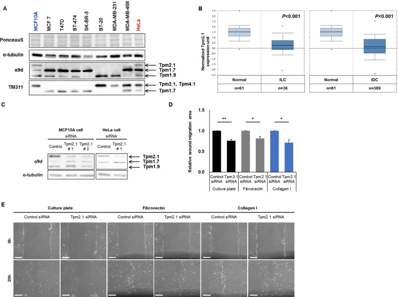
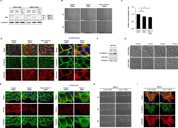
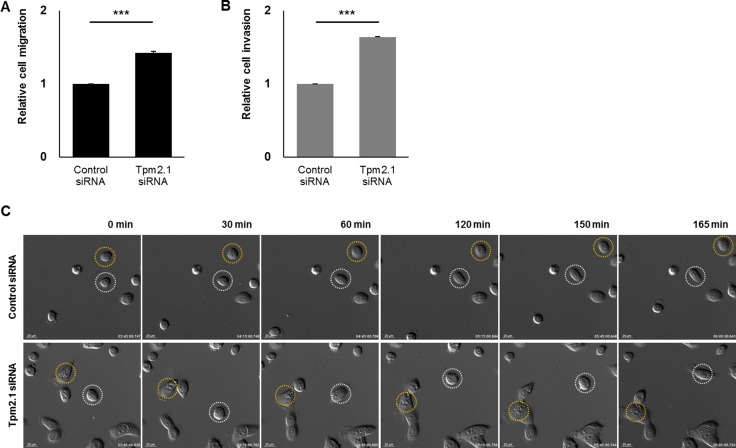
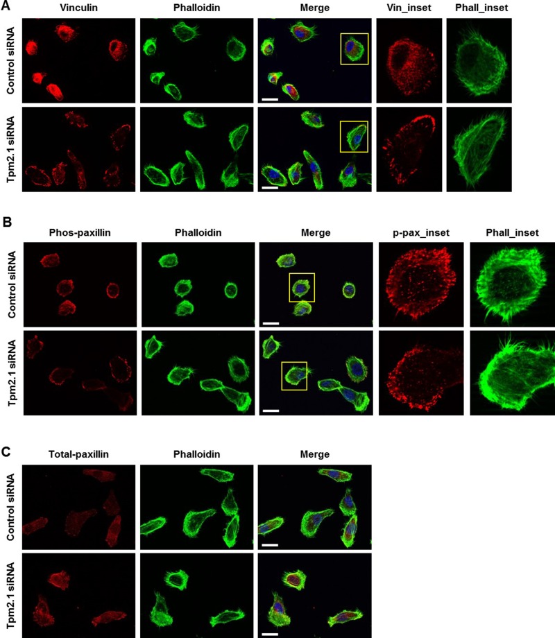
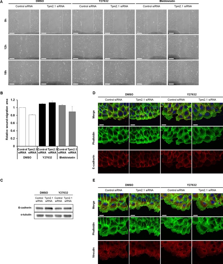
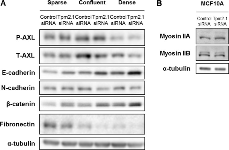
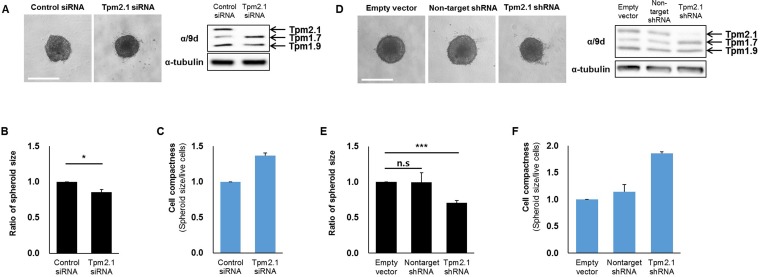
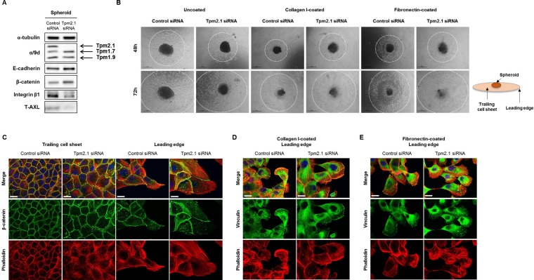
Metastasis dissemination is the result of various processes including cell migration and cell aggregation. These processes involve alterations in the expression and organization of cytoskeletal and adhesion proteins in tumor cells. Alterations in actin filaments and their binding partners are known to be key players in metastasis. Downregulation of specific tropomyosin (Tpm) isoforms is a common characteristic of transformed cells. In this study, we examined the role of Tpm2.1 in non-transformed MCF10A breast epithelial cells in cell migration and cell aggregation, because this isoform is downregulated in primary and metastatic breast cancer as well as various breast cancer cell lines. Downregulation of Tpm2.1 using siRNA or shRNA resulted in retardation of collective cell migration but increase in single cell migration and invasion. Loss of Tpm2.1 is associated with enhanced actomyosin contractility and increased expression of E-cadherin and β-catenin. Furthermore, inhibition of Rho-associated kinase (ROCK) recovered collective cell migration in Tpm2.1-silenced cells. We also found that Tpm2.1-silenced cells formed more compacted spheroids and exhibited faster cell motility when spheroids were re-plated on 2D surfaces coated with fibronectin and collagen. When Tpm2.1 was downregulated, we observed a decrease in the level of AXL receptor tyrosine kinase, which may explain the increased levels of E-cadherin and β-catenin. These studies demonstrate that Tpm2.1 functions as an important regulator of cell migration and cell aggregation in breast epithelial cells. These findings suggest that downregulation of Tpm2.1 may play a critical role during tumor progression by facilitating the metastatic potential of tumor cells.
Keywords: AXL receptor tyrosine kinase; amoeboid migration; cell aggregation; collective cell migration; metastasis.
Conflict of interest statement
CONFLICTS OF INTEREST The authors declare no conflicts of interest.
Figures
Similar articles
-
Tumor suppressor tropomyosin Tpm2.1 regulates sensitivity to apoptosis beyond anoikis characterized by changes in the levels of intrinsic apoptosis proteins.Cytoskeleton (Hoboken). 2017 Jun;74(6):233-248. doi: 10.1002/cm.21367. Epub 2017 Apr 26. Cytoskeleton (Hoboken). 2017. PMID: 28378936
-
Epigenetic silencing of TPM2 contributes to colorectal cancer progression upon RhoA activation.Tumour Biol. 2016 Sep;37(9):12477-12483. doi: 10.1007/s13277-016-5103-1. Epub 2016 Jun 23. Tumour Biol. 2016. PMID: 27333992
-
Expression of tropomyosin 2 gene isoforms in human breast cancer cell lines.Oncol Rep. 2016 Jun;35(6):3143-50. doi: 10.3892/or.2016.4732. Epub 2016 Apr 4. Oncol Rep. 2016. PMID: 27108600 Free PMC article.
-
Mechanisms of motility in metastasizing cells.Mol Cancer Res. 2010 May;8(5):629-42. doi: 10.1158/1541-7786.MCR-10-0139. Epub 2010 May 11. Mol Cancer Res. 2010. PMID: 20460404 Review.
-
Plasticity of cancer cell invasion: Patterns and mechanisms.Transl Oncol. 2021 Jan;14(1):100899. doi: 10.1016/j.tranon.2020.100899. Epub 2020 Oct 17. Transl Oncol. 2021. PMID: 33080522 Free PMC article. Review.
Cited by
-
Research Advances in the Role of the Tropomyosin Family in Cancer.Int J Mol Sci. 2023 Aug 27;24(17):13295. doi: 10.3390/ijms241713295. Int J Mol Sci. 2023. PMID: 37686101 Free PMC article. Review.
-
Local contractions regulate E-cadherin rigidity sensing.Sci Adv. 2022 Jan 28;8(4):eabk0387. doi: 10.1126/sciadv.abk0387. Epub 2022 Jan 28. Sci Adv. 2022. PMID: 35089785 Free PMC article.
-
Tropomyosin 2 exerts anti-tumor effects in lung adenocarcinoma and is a novel prognostic biomarker.Histol Histopathol. 2023 Jun;38(6):669-680. doi: 10.14670/HH-18-514. Epub 2022 Jun 14. Histol Histopathol. 2023. PMID: 36102257
-
Congenital myopathies: pathophysiological mechanisms and promising therapies.J Transl Med. 2024 Sep 2;22(1):815. doi: 10.1186/s12967-024-05626-5. J Transl Med. 2024. PMID: 39223631 Free PMC article. Review.
-
Biomarkers of tumor invasiveness in proteomics (Review).Int J Oncol. 2020 Aug;57(2):409-432. doi: 10.3892/ijo.2020.5075. Epub 2020 May 28. Int J Oncol. 2020. PMID: 32468071 Free PMC article.
References
LinkOut - more resources
Full Text Sources
Other Literature Sources
Research Materials
Miscellaneous
