. 2017 Sep 8;8(56):95481-95494.
doi: 10.18632/oncotarget.20751. eCollection 2017 Nov 10.

D-4F increases microRNA-124a and reduces neuroinflammation in diabetic stroke rats

Affiliations

D-4F increases microRNA-124a and reduces neuroinflammation in diabetic stroke rats

Ruizhuo Ning et al. Oncotarget. .

Abstract

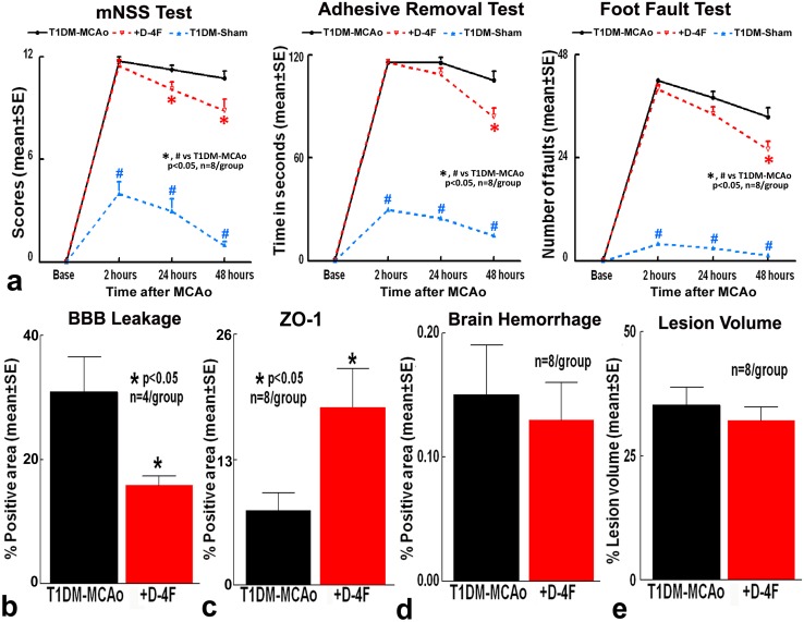
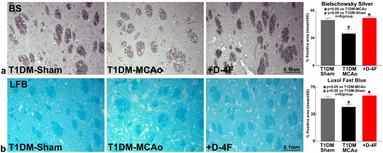
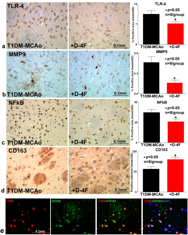
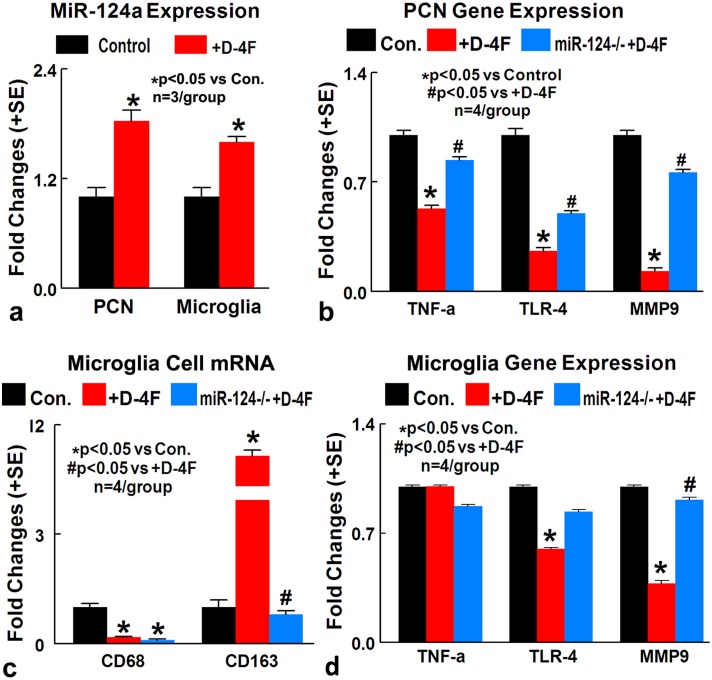
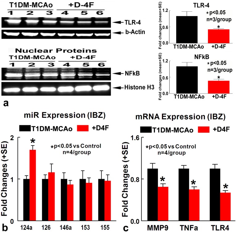
D-4F is an apolipoprotein-A1 mimetic peptide that promotes anti-inflammatory effects. MicroRNA-124 is the most abundant brain-specific microRNA and has anti-inflammatory effects. In this study, we investigated the therapeutic efficacy and mechanisms of D-4F treatment of stroke in type one diabetes mellitus (T1DM) rats. Male Wistar rats were induced with T1DM, subjected to embolic middle cerebral artery occlusion and treated with PBS or D-4F (1 mg/kg i.p.) at 2, 24 and 48 hours after stroke (n=8/group). A battery of function tests, brain blood barrier (BBB) integrity, white matter changes and microRNA expression were evaluated in vivo and in vitro. D-4F treatment in T1DM-stroke rats significantly improves functional outcome, decreases BBB leakage, increases tight junction protein expression, decreases white matter damage and inflammatory factor expression, while increasing anti-inflammatory M2 macrophage polarization in the ischemic brain. D-4F significantly increases microRNA-124a expression, and decreases matrix metalloproteinase-9, tumor necrosis factor-α and toll-like receptor-4 gene expression in the ischemic brain, and in primary cortical neuronal and microglial cultures. Inhibition of microRNA-124 in cultured primary cortical neurons and microglia attenuates D-4F induced anti-inflammatory effects and M2 macrophage polarization. D-4F treatment of T1DM-stroke increases microRNA-124 expression, promotes anti-inflammatory effects and M2 macrophage polarization, which may contribute to D-4F-induced improvement in neurological function, and BBB and white matter integrity.

Keywords: apolipoprotein-A1; diabetes mellitus; microRNA-124a; neuroinflammation; stroke.


Conflict of interest statement

CONFLICTS OF INTEREST The authors have no conflicts of interest to declare.

Figures

Figure 1
Figure 1. Stroke treatment using D-4F in T1DM rats does not decrease lesion volume and brain hemorrhagic transformation, but significantly decreases BBB leakage and improves functional outcome
Stroke in T1DM rats induces significant neurological deficits compared to T1DM-sham control rats (#p<0.05; n=8/group). D-4F treatment in T1DM stroke rats significantly: (a) improves neurological functional outcome, (b) decreases BBB leakage, and (c) increases tight junction protein Zona-occluden-1 (ZO-1) expression around blood vessels, without significantly affecting (d) brain hemorrhage and (e) lesion volume compared to PBS treated T1DM stroke rats (*p<0.05, n=8/group) at 48 hours after stroke. Data are represented as mean ± SE.
Figure 2
Figure 2. D-4F treatment of stroke in T1DM rats decreases white matter damage
Stroke in T1DM rats significantly decreases axon (Bielschowsky silver staining) and myelin density (Luxol fast blue staining) compared to T1DM-sham control rats (#p<0.05; n=8/group). D-4F treatment of stroke in T1DM rats significantly increases (a) axon density (Bielschowsky silver staining) and (b) myelin density (Luxol fast blue staining) in the ischemic border zone compared to PBS treated T1DM stroke control rats (*p<0.05, n=8/group) at 48 hours after stroke. Data are represented as mean ± SE.
Figure 3
Figure 3. D-4F treatment of stroke in T1DM rats significantly decreases inflammatory factor expression and promotes M2 macrophage polarization in the ischemic brain
At 48 hours after stroke, D-4F treatment significantly decreases (a) TLR4, (b) MMP9 and (c) NFκB expression compared to PBS treated T1DM stroke control rats (*p<0.05, n=8/group). (d) D-4F treatment also significantly promotes M2 macrophage polarization which is identified by increased CD163 expression compared to PBS treated T1DM stroke control rats (*p<0.05, n=8/group). Data are represented as mean ± SE. (e) CD163 co-localizes with CD68 indicating that CD163 is expressed by macrophages/microglia. DAPI is a nuclear stain.
Figure 5
Figure 5. In vitro, D-4F decreases inflammatory factor expression in cultured PCN and promotes M2 macrophage polarization in cultured microglia cells under conditions of high glucose and OGD
(a) D-4F treatment significantly increases miR-124a gene expression in cultured PCN and microglial cell under conditions of high glucose and OGD, compared to non-treatment control (*p<0.05, n=3/group). (b) MiR-124a inhibition in PCN cells significantly attenuates D-4F induced anti-inflammatory effects identified by significant increases in TNFα, MMP9 and TLR4 gene expression compared to D-4F treated PCN (*p<0.05 vs. control, #p<0.05 vs. D-4F treatment, n=4/group). (c) MiR-124 knock down in microglia culture significantly attenuates D-4F induced M2-macrophage polarization (*p<0.05 vs. control, #p<0.05 vs. D-4F treatment, n=4/group). (d) MiR-124a inhibition in microglial cells significantly attenuates D-4F induced anti-inflammatory effects identified by increases in MMP9 (*p<0.05 vs. control, #p<0.05 vs. D-4F treatment, n=4/group) and TLR4 gene expression compared to D-4F treated microglia. Data are represented as mean ± SE.
Figure 4
Figure 4. D-4F treatment of stroke in T1DM rats significantly decreases inflammatory factor expression and increases miR-124a expression
(a) Western blot assay showing that D-4F treatment of T1DM stroke rats decreases TLR4, and nuclear NFκB expression in the ischemic brain compared to PBS treated T1DM stroke control rats (*p<0.05, n=3/group). (b) D-4F treatment significantly increases miR-124a expression in the ischemic brain at 48 hours after stroke (*p<0.05, n=4/group). (c) D-4F treatment significantly decreases MMP9, TLR4 and TNFα gene expression in the ischemic brain compared to PBS treated T1DM stroke control rats at 48 hours after stroke (*p<0.05, n=4/group). Data are represented as mean ± SE.

References

    1. Putaala J, Liebkind R, Gordin D, Thorn LM, Haapaniemi E, Forsblom C, Groop PH, Kaste M, Tatlisumak T. Diabetes mellitus and ischemic stroke in the young: clinical features and long-term prognosis. Neurology. 2011;76:1831–7. https://doi.org/10.1212/WNL.0b013e31821cccc2. - DOI - PubMed
    1. Yan T, Venkat P, Ye X, Chopp M, Zacharek A, Ning R, Cui Y, Roberts C, Kuzmin-Nichols N, Sanberg CD, Chen J. HUCBCs increase angiopoietin 1 and induce neurorestorative effects after stroke in T1DM rats. CNS Neurosci Ther. 2014;20:935–44. https://doi.org/10.1111/cns.12307. - DOI - PMC - PubMed
    1. Ning R, Chopp M, Yan T, Zacharek A, Zhang C, Roberts C, Cui X, Lu M, Chen J. Tissue plasminogen activator treatment of stroke in type-1 diabetes rats. Neuroscience. 2012;222:326–32. https://doi.org/10.1016/j.neuroscience.2012.07.018. - DOI - PMC - PubMed
    1. Ning R, Chopp M, Zacharek A, Yan T, Zhang C, Roberts C, Lu M, Chen J. Neamine induces neuroprotection after acute ischemic stroke in type one diabetic rats. Neuroscience. 2014;257:76–85. https://doi.org/10.1016/j.neuroscience.2013.10.071. - DOI - PMC - PubMed
    1. Fan X, Qiu J, Yu Z, Dai H, Singhal AB, Lo EH, Wang X. A rat model of studying tissue-type plasminogen activator thrombolysis in ischemic stroke with diabetes. Stroke. 2012;43:567–70. https://doi.org/10.1161/strokeaha.111.635250. - DOI - PMC - PubMed

LinkOut - more resources