

CHST6 mutation screening and endoplasmatic reticulum stress in macular corneal dystrophy
- PMID: 29221207
- PMCID: PMC5707101
- DOI: 10.18632/oncotarget.22028
CHST6 mutation screening and endoplasmatic reticulum stress in macular corneal dystrophy
Abstract
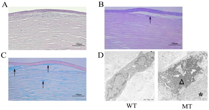
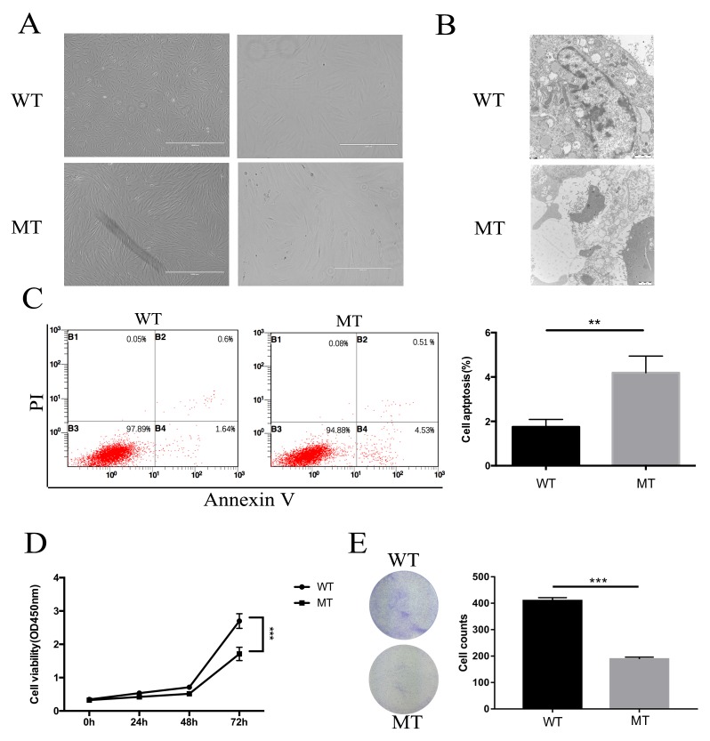
Macular corneal dystrophy (MCD) is an autosomal recessive disorder mainly caused by gene mutations of carbohydrate sulfotransferase (CHST6) leading to bilateral visual impairment. Because the mechanism underlying this degeneration remains poorly understood, we investigated molecular alterations and pathways that may be involved in MCD in this issue. Different mutation sites were screened by direct sequencing of the coding region of CHST6. In addition, we described morphological changes in MCD keratocytes by light microscopy and electron microscopy and determined the relationship between the development of this disease and the occurrence of apoptosis through flow cytometry, cell counting kit-8, colony formation assay and other experiments. Western blotting and quantitative real-time polymerase chain reaction were used to determine if endoplasmic reticulum (ER) stress was activated. We found 10 kinds of mutations among these families with 3 novel mutations included. The percentage of apoptotic keratocytes increased in MCD patients; furthermore, the expression of apoptosis related protein B-cell lymphoma-2 (Bcl-2) was down-regulated while Bcl-2 associated X protein was upregulated. Finally, ER stress was activated with the upregulation of glucose-regulated protein 78 and CCAAT-enhancer-binding protein homologous protein. Our clinical and in vitro results suggest that the CHST6 mutation associated with MCD is associated with apoptosis, and ER stress is probably involved in this apoptosis pathway.
Keywords: CHST6; apoptosis; endoplasmic reticulum stress; keratocytes; macular corneal dystrophy.
Conflict of interest statement
CONFLICTS OF INTEREST The authors declare no conflicts of interest.
Figures


Similar articles
-
Association of macular corneal dystrophy with excessive cell senescence and apoptosis induced by the novel mutant CHST6.Exp Eye Res. 2022 Jan;214:108862. doi: 10.1016/j.exer.2021.108862. Epub 2021 Nov 24. Exp Eye Res. 2022. PMID: 34826417
-
Identification of novel mutations in the carbohydrate sulfotransferase gene (CHST6) causing macular corneal dystrophy.Invest Ophthalmol Vis Sci. 2002 Feb;43(2):377-82. Invest Ophthalmol Vis Sci. 2002. PMID: 11818380
-
Novel mutations in the CHST6 gene causing macular corneal dystrophy.Clin Genet. 2004 Feb;65(2):120-5. doi: 10.1111/j.0009-9163.2004.00191.x. Clin Genet. 2004. PMID: 14984470
-
Bilateral phototherapeutic keratectomy for corneal macular dystrophy in an adolescent: case report and review of the literature.Ophthalmic Genet. 2020 Aug;41(4):368-372. doi: 10.1080/13816810.2020.1776335. Epub 2020 Jun 16. Ophthalmic Genet. 2020. PMID: 32543930 Review.
-
Macular Corneal Dystrophy: An Updated Review.Curr Eye Res. 2021 Jun;46(6):765-770. doi: 10.1080/02713683.2020.1849727. Epub 2020 Nov 29. Curr Eye Res. 2021. PMID: 33171054 Review.
Cited by
-
Trehalose extricates impaired mitochondrial and autophagy dysregulation in patient iPSC-derived macular corneal dystrophy disease model.Stem Cell Res Ther. 2024 Dec 5;15(1):464. doi: 10.1186/s13287-024-04016-4. Stem Cell Res Ther. 2024. PMID: 39639354 Free PMC article.
-
Novel Mutations in COL6A3 That Associated With Peters' Anomaly Caused Abnormal Intracellular Protein Retention and Decreased Cellular Resistance to Oxidative Stress.Front Cell Dev Biol. 2020 Nov 10;8:531986. doi: 10.3389/fcell.2020.531986. eCollection 2020. Front Cell Dev Biol. 2020. PMID: 33304895 Free PMC article.
-
Endoplasmic reticulum stress: molecular mechanism and therapeutic targets.Signal Transduct Target Ther. 2023 Sep 15;8(1):352. doi: 10.1038/s41392-023-01570-w. Signal Transduct Target Ther. 2023. PMID: 37709773 Free PMC article. Review.
-
Matrix glycosaminoglycans and proteoglycans in human cornea organoids and similarities with fetal corneal stages.Ocul Surf. 2025 Jan;35:68-80. doi: 10.1016/j.jtos.2024.11.007. Epub 2024 Nov 28. Ocul Surf. 2025. PMID: 39615587
-
Genetic implications of CHST6 gene mutations and their corneal microstructural changes in macular corneal dystrophy patients.Mol Vis. 2024 Oct 4;30:305-318. eCollection 2024. Mol Vis. 2024. PMID: 39959172 Free PMC article.
References
-
- Midura RJ, Hascall VC, Maccallum DK, Meyer RF, Thonar EJ, Hassell JR, Smith CF, Klintworth GK. Proteoglycan biosynthesis by human corneas from patients with types 1 and 2 macular corneal dystrophy. J Biol Chem. 1990;265:15947–55. - PubMed
-
- Klintworth GK, Oshima E, Al-Rajhi A, Al-Saif A, Thonar EJ, Karcioglu ZA. Macular corneal dystrophy in Saudi Arabia: a study of 56 cases and recognition of a new immunophenotype. Am J Ophthalmol. 1998;125:417. - PubMed
-
- Edward DP, Thonar EJ, Srinivasan M, Yue BJ, Tso MO. Macular dystrophy of the cornea. A systemic disorder of keratan sulfate metabolism. Ophthalmology. 1990;97:1194–200. - PubMed
LinkOut - more resources
Full Text Sources
Other Literature Sources
