

PURA, the gene encoding Pur-alpha, member of an ancient nucleic acid-binding protein family with mammalian neurological functions
- PMID: 29221753
- PMCID: PMC5770235
- DOI: 10.1016/j.gene.2017.12.004
PURA, the gene encoding Pur-alpha, member of an ancient nucleic acid-binding protein family with mammalian neurological functions
Abstract
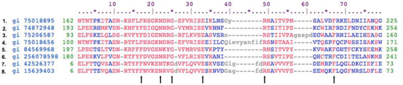
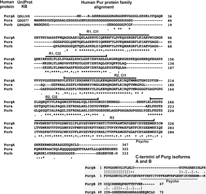
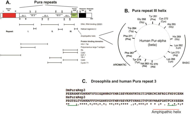
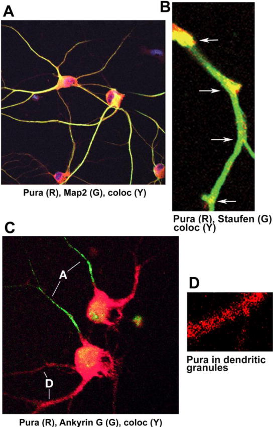
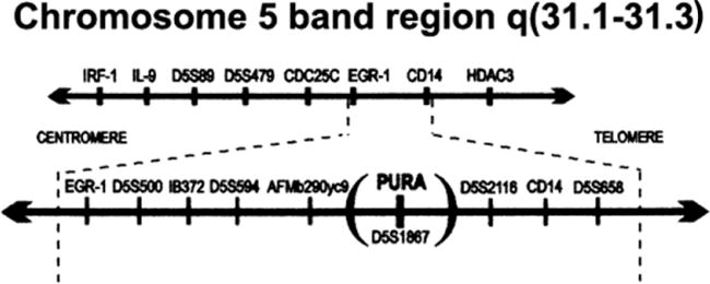
The PURA gene encodes Pur-alpha, a 322 amino acid protein with repeated nucleic acid binding domains that are highly conserved from bacteria through humans. PUR genes with a single copy of this domain have been detected so far in spirochetes and bacteroides. Lower eukaryotes possess one copy of the PUR gene, whereas chordates possess 1 to 4 PUR family members. Human PUR genes encode Pur-alpha (Pura), Pur-beta (Purb) and two forms of Pur-gamma (Purg). Pur-alpha is a protein that binds specific DNA and RNA sequence elements. Human PURA, located at chromosome band 5q31, is under complex control of three promoters. The entire protein coding sequence of PURA is contiguous within a single exon. Several studies have found that overexpression or microinjection of Pura inhibits anchorage-independent growth of oncogenically transformed cells and blocks proliferation at either G1-S or G2-M checkpoints. Effects on the cell cycle may be mediated by interaction of Pura with cellular proteins including Cyclin/Cdk complexes and the Rb tumor suppressor protein. PURA knockout mice die shortly after birth with effects on brain and hematopoietic development. In humans environmentally induced heterozygous deletions of PURA have been implicated in forms of myelodysplastic syndrome and progression to acute myelogenous leukemia. Pura plays a role in AIDS through association with the HIV-1 protein, Tat. In the brain Tat and Pura association in glial cells activates transcription and replication of JC polyomavirus, the agent causing the demyelination disease, progressive multifocal leukoencephalopathy. Tat and Pura also act to stimulate replication of the HIV-1 RNA genome. In neurons Pura accompanies mRNA transcripts to sites of translation in dendrites. Microdeletions in the PURA locus have been implicated in several neurological disorders. De novo PURA mutations have been related to a spectrum of phenotypes indicating a potential PURA syndrome. The nucleic acid, G-rich Pura binding element is amplified as expanded polynucleotide repeats in several brain diseases including fragile X syndrome and a familial form of amyotrophic lateral sclerosis/fronto-temporal dementia. Throughout evolution the Pura protein plays a critical role in survival, based on conservation of its nucleic acid binding properties. These Pura properties have been adapted in higher organisms to the as yet unfathomable development of the human brain.
Keywords: AIDS; ALS; AML; Acute myelogenous leukemia; Amyotrophic lateral sclerosis; C9ORF72; Dementia; FMR1; FXS; Fragile X syndrome; JCV; Myelodysplastic syndrome; PML; Polyomavirus JC; Progressive multifocal leukoencephalopathy; Pur-beta; Pur-gamma-A and Pur-gamma-B; Pura (Pur-alpha); Purb; Purg.
Copyright © 2017 Elsevier B.V. All rights reserved.
Figures
Similar articles
-
Deletions of PURA, at 5q31, and PURB, at 7p13, in myelodysplastic syndrome and progression to acute myelogenous leukemia.Leukemia. 2001 Jun;15(6):954-62. doi: 10.1038/sj.leu.2402108. Leukemia. 2001. PMID: 11417483
-
Activation of the JC virus Tat-responsive transcriptional control element by association of the Tat protein of human immunodeficiency virus 1 with cellular protein Pur alpha.Proc Natl Acad Sci U S A. 1996 Nov 26;93(24):14112-7. doi: 10.1073/pnas.93.24.14112. Proc Natl Acad Sci U S A. 1996. PMID: 8943069 Free PMC article.
-
A synthetic Pur-based peptide binds and alters G-quadruplex secondary structure present in the expanded RNA repeat of C9orf72 ALS/FTD.Biochim Biophys Acta Mol Cell Res. 2020 Jun;1867(6):118674. doi: 10.1016/j.bbamcr.2020.118674. Epub 2020 Feb 6. Biochim Biophys Acta Mol Cell Res. 2020. PMID: 32035967 Free PMC article.
-
Multiple roles for Puralpha in cellular and viral regulation.Cell Cycle. 2009 Feb 1;8(3):1-7. doi: 10.4161/cc.8.3.7585. Epub 2009 Feb 10. Cell Cycle. 2009. PMID: 19182532 Free PMC article. Review.
-
The Pur protein family: clues to function from recent studies on cancer and AIDS.Anticancer Res. 2003 May-Jun;23(3A):2093-100. Anticancer Res. 2003. PMID: 12894583 Review.
Cited by
-
PURA syndrome-a genetic cause of a neurodevelopmental disorder-case report.Front Pediatr. 2025 Jul 10;13:1607213. doi: 10.3389/fped.2025.1607213. eCollection 2025. Front Pediatr. 2025. PMID: 40708900 Free PMC article.
-
Regulatory mechanism of circular RNAs in neurodegenerative diseases.CNS Neurosci Ther. 2024 Apr;30(4):e14499. doi: 10.1111/cns.14499. Epub 2023 Oct 21. CNS Neurosci Ther. 2024. PMID: 37864389 Free PMC article. Review.
-
PURα Promotes the Transcriptional Activation of PCK2 in Oesophageal Squamous Cell Carcinoma Cells.Genes (Basel). 2020 Oct 31;11(11):1301. doi: 10.3390/genes11111301. Genes (Basel). 2020. PMID: 33142842 Free PMC article.
-
Neonatal PURA syndrome: a case report and literature review.Transl Pediatr. 2021 Jan;10(1):194-203. doi: 10.21037/tp-20-248. Transl Pediatr. 2021. PMID: 33633953 Free PMC article.
-
Incorporating mutational heterogeneity to identify genes that are enriched for synonymous mutations in cancer.BMC Bioinformatics. 2023 Dec 7;24(1):462. doi: 10.1186/s12859-023-05521-8. BMC Bioinformatics. 2023. PMID: 38062391 Free PMC article.
References
-
- Andres AM, Soldevila M, Lao O, Volpini V, Saitou N, Jacobs HT, Hayasaka I, Calafell F, Bertranpetit J. Comparative genetics of functional trinucleotide tandem repeats in humans and apes. J Mol Evol. 2004;59:329–39. - PubMed
-
- Ash PE, Bieniek KF, Gendron TF, Caulfield T, Lin WL, Dejesus-Hernandez M, van Blitterswijk MM, Jansen-West K, Paul JW, 3rd, Rademakers R, Boylan KB, Dickson DW, Petrucelli L. Unconventional translation of C9ORF72 GGGGCC expansion generates insoluble polypeptides specific to c9FTD/ALS. Neuron. 2013;77:639–46. - PMC - PubMed
-
- Aumiller V, Graebsch A, Kremmer E, Niessing D, Forstemann K. Drosophila Pur-alpha binds to trinucleotide-repeat containing cellular RNAs and translocates to the early oocyte. RNA Biol. 2012;9:633–43. - PubMed
-
- Barr SM, Johnson EM. Ras-induced colony formation and anchorage-independent growth inhibited by elevated expression of Puralpha in NIH3T3 cells. J Cell Biochem. 2001;81:621–38. - PubMed
Publication types
MeSH terms
Substances
Grants and funding
LinkOut - more resources
Full Text Sources
Other Literature Sources
Miscellaneous
