. 2018 Feb 7;26(2):634-647.
doi: 10.1016/j.ymthe.2017.11.008. Epub 2017 Nov 16.

The Impact of the CD9 Tetraspanin on Lentivirus Infectivity and Exosome Secretion

Affiliations

The Impact of the CD9 Tetraspanin on Lentivirus Infectivity and Exosome Secretion

Kai O Böker et al. Mol Ther. .

Abstract

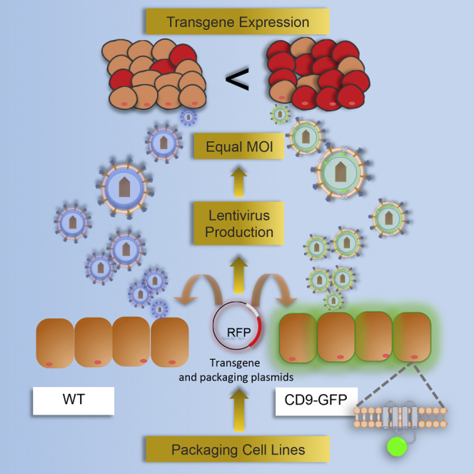
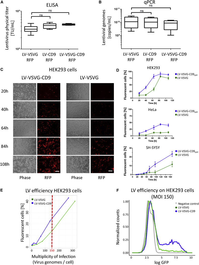
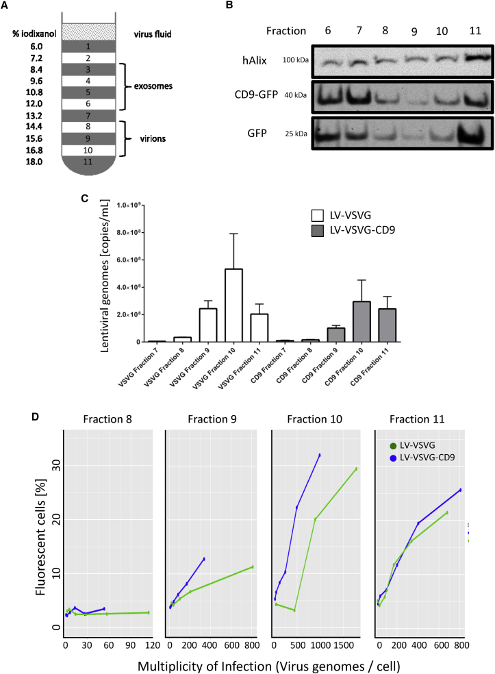
Efficient transduction tools are a hallmark for both research and therapy development. Here, we introduce new insights into the generation of lentiviral vectors with improved performance by utilizing producer cells with increased production rates of extracellular vesicles through CD9 overexpression. Most human cells secrete small vesicles from their surface (microvesicles) or intraluminal endosome-derived membranes (exosomes). In particular, enhanced levels of the tetraspanin CD9 result in significantly increased numbers of extracellular vesicles with exosome-like features that were secreted from four different human cell lines. Intriguingly, exosomes and their biogenesis route display similarities to lentivirus and we examined the impact of CD9 expression on release and infectivity of recombinant lentiviral vectors. Although the titers of released viral particles were not increased upon production in high CD9 cells, we observed improved performance in terms of both speed and efficiency of lentiviral gene delivery into numerous human cell lines, including HEK293, HeLa, SH-SY5Y, as well as B and T lymphocytes. Here, we demonstrate that enhanced CD9 enables lentiviral transduction in the absence of any pseudotyping viral glycoprotein or fusogenic molecule. Our findings indicate an important role of CD9 for lentiviral vector and exosome biogenesis and point out a remarkable function of this tetraspanin in membrane fusion, viral infectivity, and exosome-mediated horizontal information transfer.

Keywords: CD9; exosomes; extracellular vesicles; lentivirus; tetraspanin.


Figures

None
Graphical abstract
Figure 1
Figure 1
Overexpression of Exosomal Marker Proteins CD9, Alix, and TSG101 in HEK293 Cells Affects Vesicle Amounts and Sizes (A) qPCR analyses were performed and displayed a 4- to 22-fold higher expression of the respective transgenes. Nanoparticle tracking analysis of extracellular vesicles showed significantly enlarged particles for HEK293-TSG101 and HEK293-Alix compared to wild-type. (B) The overexpression of CD9 led to a decreased average size of secreted vesicles. (C) Total extracellular vesicle amount was decreased upon TSG101 and Alix overexpression, but the extracellular vesicle amount was significantly increased upon CD9 overexpression. (D) For vesicles within the size range of exosomes (30–100 nm), these trends were even more obvious. Error bars represent SD.
Figure 2
Figure 2
CD9 Locates to the Plasma Membrane and Cell-Cell Contact Surfaces in Stably CD9-GFP-Expressing Cell Lines Cell membrane localization of CD9-GFP was observed via fluorescence microscopy. Green fluorescence was detected after at least 2 weeks of Blasticidin selection (scale bar, 100 μm).
Figure 3
Figure 3
Lentivirus Production and Comparison between Exosomes and LVs (A) HEK293FT or HEK293FT-CD9 cells were transfected with three different plasmids encoding for envelope glycoproteins (i.e., CD9 and/or VSVG), viral capsid proteins, and the gene of interest, here, an RFP. LVs are shed from the cell membrane, and LVs produced in HEK293FT-CD9 cells carry additional CD9-GFP and/or VSVG within their envelope. (B) The three different LVs used in this study, i.e., exposing VSVG, CD9, or both VSVG and CD9, are schematically shown.
Figure 4
Figure 4
Production Capacity and Transduction Efficiency of LV-VSVG and LV-VSVG-CD9 (A and B) Comparative analysis of three lentiviral productions revealed only minor differences in titer concentrations for LV-VSVG, LV-VSVG-CD9GFP, and LV-CD9GFP demonstrated by ELISA (A) and qPCR (B). Comparison of transduction efficiency was evaluated with LV-VSVG as a standard control and LV-VSVG-CD9GF on HEK293, HeLa, and SH-SY5Y cells. (C) Successful transduction was confirmed via fluorescence microscopy in regular intervals from 20 to 108 hr after transduction. (D) In fluorescence microscopy analysis, positive cells were quantified by manual counting and the transduction efficiency was calculated. (E) Comparing equal amounts of LV-VSVG and LV-VSVG-CD9 revealed an increased efficiency for LV-VSVG-CD9 over a broad MOI range (30–300 MOI). (F) Density plot representation of lentiviral efficiency at equal MOI (150) through FACS analysis. Scale bars, 200 μm. Error bars represent SD.
Figure 5
Figure 5
CD9 Mediates Fusion with Target Cell Membrane (A) HEK293 cells were transduced with CD9, VSVG, and VSVG-CD9 LVs. All tested LVs show expression after 72 hr. The highest efficiency was observed with LV-VSVG-CD9GFP, followed by LV-VSVG, but the virus without any viral glycoprotein (LV-CD9GFP) also successfully infected a minor proportion of cells. (B) Negative control without viral envelope proteins or CD9GFP shows no transduction. Scale bars, 100 μm (A) and 200 μm (B). Exposure time: 1,000 ms.
Figure 6
Figure 6
Density Gradient Centrifugation Separates Exosomes and Lentiviral Particles (A) Lentiviral supernatant was fractionated by centrifugation through a gradient of iodixanol to separate lentiviral particles and exosomes. (B) Western blot analysis revealed high concentrations of CD9-GFP and GFP in fraction 6+7 and 10+11, whereas Alix showed an equal distribution, indicating a successful separation of exosomes (fraction 6+7) and lentiviruses (fraction 10+11). (C) qPCR titer measurements clarified the presence of lentiviral particles in fractions 9–11. (D) Comparative analysis of LV-VSVG and LV-VSVG-CD9 with similar MOIs displayed no enhanced infectivity for fractions 8 and 11, whereas in fractions 9 and 11, increased transduction rates were observed for LV-VSVG-CD9 lentiviral constructs on HEK293 cells. Error bars represent SD.

Similar articles

Cited by

References

    1. Anderson J.L., Hope T.J. Intracellular trafficking of retroviral vectors: obstacles and advances. Gene Ther. 2005;12:1667–1678. - PubMed
    1. Bukrinsky M.I., Haggerty S., Dempsey M.P., Sharova N., Adzhubel A., Spitz L., Lewis P., Goldfarb D., Emerman M., Stevenson M. A nuclear localization signal within HIV-1 matrix protein that governs infection of non-dividing cells. Nature. 1993;365:666–669. - PMC - PubMed
    1. Dull T., Zufferey R., Kelly M., Mandel R.J., Nguyen M., Trono D., Naldini L. A third-generation lentivirus vector with a conditional packaging system. J. Virol. 1998;72:8463–8471. - PMC - PubMed
    1. Zufferey R., Dull T., Mandel R.J., Bukovsky A., Quiroz D., Naldini L., Trono D. Self-inactivating lentivirus vector for safe and efficient in vivo gene delivery. J. Virol. 1998;72:9873–9880. - PMC - PubMed
    1. Burns J.C., Friedmann T., Driever W., Burrascano M., Yee J.K. Vesicular stomatitis virus G glycoprotein pseudotyped retroviral vectors: concentration to very high titer and efficient gene transfer into mammalian and nonmammalian cells. Proc. Natl. Acad. Sci. USA. 1993;90:8033–8037. - PMC - PubMed

Publication types