

Skeletal Muscle Regenerative Potential of Human MuStem Cells following Transplantation into Injured Mice Muscle
- PMID: 29221805
- PMCID: PMC5835152
- DOI: 10.1016/j.ymthe.2017.10.013
Skeletal Muscle Regenerative Potential of Human MuStem Cells following Transplantation into Injured Mice Muscle
Abstract
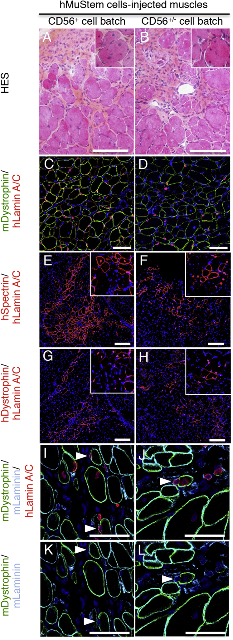
After intra-arterial delivery in the dystrophic dog, allogeneic muscle-derived stem cells, termed MuStem cells, contribute to long-term stabilization of the clinical status and preservation of the muscle regenerative process. However, it remains unknown whether the human counterpart could be identified, considering recent demonstrations of divergent features between species for several somatic stem cells. Here, we report that MuStem cells reside in human skeletal muscle and display a long-term ability to proliferate, allowing generation of a clinically relevant amount of cells. Cultured human MuStem (hMuStem) cells do not express hematopoietic, endothelial, or myo-endothelial cell markers and reproducibly correspond to a population of early myogenic-committed progenitors with a perivascular/mesenchymal phenotypic signature, revealing a blood vessel wall origin. Importantly, they exhibit both myogenesis in vitro and skeletal muscle regeneration after intramuscular delivery into immunodeficient host mice. Together, our findings provide new insights supporting the notion that hMuStem cells could represent an interesting therapeutic candidate for dystrophic patients.
Keywords: DMD; MuStem; cell therapy; human adult stem cells; regenerative medicine; skeletal muscle.
Copyright © 2017 The American Society of Gene and Cell Therapy. Published by Elsevier Inc. All rights reserved.
Figures
Similar articles
-
Systemic delivery of allogenic muscle stem cells induces long-term muscle repair and clinical efficacy in duchenne muscular dystrophy dogs.Am J Pathol. 2011 Nov;179(5):2501-18. doi: 10.1016/j.ajpath.2011.07.022. Epub 2011 Sep 13. Am J Pathol. 2011. PMID: 21924229 Free PMC article.
-
Differential Gene Expression Profiling of Dystrophic Dog Muscle after MuStem Cell Transplantation.PLoS One. 2015 May 8;10(5):e0123336. doi: 10.1371/journal.pone.0123336. eCollection 2015. PLoS One. 2015. PMID: 25955839 Free PMC article.
-
Identification in GRMD dog muscle of critical miRNAs involved in pathophysiology and effects associated with MuStem cell transplantation.BMC Musculoskelet Disord. 2016 May 11;17:209. doi: 10.1186/s12891-016-1060-5. BMC Musculoskelet Disord. 2016. PMID: 27170302 Free PMC article.
-
Initial failure in myoblast transplantation therapy has led the way toward the isolation of muscle stem cells: potential for tissue regeneration.Curr Top Dev Biol. 2005;68:263-80. doi: 10.1016/S0070-2153(05)68009-X. Curr Top Dev Biol. 2005. PMID: 16125002 Review.
-
Insights into skeletal muscle development and applications in regenerative medicine.Int Rev Cell Mol Biol. 2013;300:51-83. doi: 10.1016/B978-0-12-405210-9.00002-3. Int Rev Cell Mol Biol. 2013. PMID: 23273859 Review.
Cited by
-
Characterization of traumatized muscle-derived multipotent progenitor cells from low-energy trauma.Stem Cell Res Ther. 2021 Jan 6;12(1):6. doi: 10.1186/s13287-020-02038-2. Stem Cell Res Ther. 2021. PMID: 33407850 Free PMC article.
-
The First Comprehensive Cohort of the Duchenne Muscular Dystrophy in Iranian Population: Mutation Spectrum of 314 Patients and Identifying Two Novel Nonsense Mutations.J Mol Neurosci. 2020 Oct;70(10):1565-1573. doi: 10.1007/s12031-020-01594-9. Epub 2020 May 21. J Mol Neurosci. 2020. PMID: 32436198
-
Duchenne's Muscular Dystrophy: The Role of Induced Pluripotent Stem Cells and Genomic Editing on Muscle Regeneration.Cureus. 2020 Sep 22;12(9):e10600. doi: 10.7759/cureus.10600. Cureus. 2020. PMID: 33123420 Free PMC article. Review.
-
Human MuStem cells repress T-cell proliferation and cytotoxicity through both paracrine and contact-dependent pathways.Stem Cell Res Ther. 2022 Jan 10;13(1):7. doi: 10.1186/s13287-021-02681-3. Stem Cell Res Ther. 2022. PMID: 35012660 Free PMC article.
-
Human MuStem Cell Grafting into Infarcted Rat Heart Attenuates Adverse Tissue Remodeling and Preserves Cardiac Function.Mol Ther Methods Clin Dev. 2020 Jun 15;18:446-463. doi: 10.1016/j.omtm.2020.06.009. eCollection 2020 Sep 11. Mol Ther Methods Clin Dev. 2020. PMID: 32695846 Free PMC article.
References
-
- Emery A.E.H. Population frequencies of inherited neuromuscular diseases--a world survey. Neuromuscul. Disord. 1991;1:19–29. - PubMed
-
- Hoffman E.P., Brown R.H., Jr., Kunkel L.M. Dystrophin: the protein product of the Duchenne muscular dystrophy locus. Cell. 1987;51:919–928. - PubMed
-
- Eagle M., Bourke J., Bullock R., Gibson M., Mehta J., Giddings D., Straub V., Bushby K. Managing Duchenne muscular dystrophy--the additive effect of spinal surgery and home nocturnal ventilation in improving survival. Neuromuscul. Disord. 2007;17:470–475. - PubMed
-
- Kohler M., Clarenbach C.F., Bahler C., Brack T., Russi E.W., Bloch K.E. Disability and survival in Duchenne muscular dystrophy. J. Neurol. Neurosurg. Psychiatry. 2009;80:320–325. - PubMed
Publication types
MeSH terms
LinkOut - more resources
Full Text Sources
Other Literature Sources
