. 2018 Feb 2;131(3):jcs202580.
doi: 10.1242/jcs.202580.

Blood vitronectin is a major activator of LIF and IL-6 in the brain through integrin-FAK and uPAR signaling

Affiliations

Blood vitronectin is a major activator of LIF and IL-6 in the brain through integrin-FAK and uPAR signaling

Matthew P Keasey et al. J Cell Sci. .

Abstract

We defined how blood-derived vitronectin (VTN) rapidly and potently activates leukemia inhibitory factor (LIF) and pro-inflammatory interleukin 6 (IL-6) in vitro and after vascular injury in the brain. Treatment with VTN (but not fibrinogen, fibronectin, laminin-111 or collagen-I) substantially increased LIF and IL-6 within 4 h in C6-astroglioma cells, while VTN-/- mouse plasma was less effective than that from wild-type mice. LIF and IL-6 were induced by intracerebral injection of recombinant human (rh)VTN in mice, but induction seen upon intracerebral hemorrhage was less in VTN-/- mice than in wild-type littermates. In vitro, VTN effects were inhibited by RGD, αvβ3 and αvβ5 integrin-blocking peptides and antibodies. VTN activated focal adhesion kinase (FAK; also known as PTK2), whereas pharmacological- or siRNA-mediated inhibition of FAK, but not PYK2, reduced the expression of LIF and IL-6 in C6 and endothelial cells and after traumatic cell injury. Dominant-negative FAK (Y397F) reduced the amount of injury-induced LIF and IL-6. Pharmacological inhibition or knockdown of uPAR (also known as PLAUR), which binds VTN, also reduced cytokine expression, possibly through a common target of uPAR and integrins. We propose that VTN leakage into tissues promotes inflammation. Integrin-FAK signaling is therefore a novel IL-6 and LIF regulation mechanism relevant to the inflammation and stem cell fields.

Keywords: FAK; IL-6; Integrin; LIF; Vitronectin; uPAR.


Conflict of interest statement

Competing interestsThe authors declare no competing or financial interests.

Figures

Fig. 1.
Fig. 1.
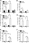
Among ECM proteins, VTN uniquely activates LIF and IL-6 expression in C6 astroglioma cells. Serum contains a variety and various abundances of integrin-binding ECM proteins. We plated C6 cells for 24 h in 10% serum, then replaced the medium with a low serum formulation (1% v/v; LS) for 24 h, which reduced LIF (A) and IL-6 (B) mRNA expression, as measured by RT-qPCR, relative to that seen in control 10% serum (Ctrl). (C) In the same experiment as in A and B, CNTF mRNA was increased, likely due to inhibitory activity of serum ECMs, such as VTN, on CNTF expression which is negatively regulated by integrin–FAK signaling. In further experiments, we seeded C6 cells and maintained them for 24 h before serum was removed for a further 24 h. Then, VTN was ‘spiked’ into the medium (10 μg/ml concentration), where it rapidly (within 4 h) and robustly induced LIF (D) and IL-6 (E) mRNA relative to what was seen upon addition of PBS control (vehicle, no ECM substrate). Fibronectin (FN1), fibrinogen (FGN), laminin-111 (LM) or collagen-I (COL) addition had no effect. VTN increased LIF (F) and IL-6 (G) in a dose-dependent fashion. NS, no serum; concentrations of VTN are μg/ml. VTN treatment for 4 h caused increased release of LIF (H) and IL-6 (I) protein in conditioned medium, as shown by a dot blot assay (representative of three independent experiments). Plasma from VTN−/− mice induced LIF (J) and IL-6 (K) gene expression by less than plasma from VTN+/+ littermates when added for 4 h to C6 cells that had been serum-deprived for 24 h. Data are means±s.e.m. of three or four independent experiments (as denoted in columns). **P<0.01, ***P<0.001.
Fig. 2.
Fig. 2.
VTN regulates LIF and IL-6 expression in mouse brain. Injection of 1 µg rhVTN into the striatum of adult VTN+/+ or VTN−/− littermate mice leads to increased LIF mRNA (A), with no induction observed with heat denatured rhVTN (B). (C) rhVTN also induced IL-6 mRNA expression at 24 h relative to that seen upon PBS injections (VTN+/+ mice: PBS n=7, VTN n=7; VTN−/− mice: PBS, n=5, VTN, n=5) with no difference seen when using heat denatured rhVTN (D). Total LIF (E) and IL-6 (F) protein were induced by rhVTN injection into the striatum of the same mice as measured by ELISA. Hemorrhagic leakage induced through collagenase injection into the striatum caused a greater increase in LIF (G) and IL-6 (H) mRNA in VTN+/+ than in VTN−/− littermate mice after 24 h (n=6–7/group). Data are means±s.e.m. *P<0.05, **P<0.01, ***P<0.001.
Fig. 3.
Fig. 3.
Integrins mediate VTN-induced LIF and IL-6 in C6 cells. C6 cells were seeded onto plastic for 24 h in serum-containing medium, then serum starved for 24 h before addition of VTN to the medium for 4 h. Co-treatment with RGD or αvβ3 (P11) integrin-blocking peptides reduced the effects of VTN on LIF (A) and IL-6 (B) mRNA expression. RAD is a non-RGD control peptide. LIF and IL-6 expression were not completely abolished in these experiments, suggesting that there is an additional VTN-activated mechanism. Indeed, BC-11, a uPA–uPAR inhibitor, decreased VTN-induced LIF (C) and IL-6 (D) mRNA expression, but did not have any effect alone, after 4 h. Data are means±s.e.m. of four independent experiments. Since 1 µg/ml VTN was effective in inducing LIF and IL-6, we only chose this dosage for these experiments. Co-incubation of both P11 and BC-11 could not further decrease the VTN-mediated LIF and IL-6 induction. Finally, knockdown of uPAR by means of siRNA (siUPAR) reduced by LIF (E) and IL-6 (F) induction by VTN (10 µg/ml). uPAR knockdown was confirmed by RT-qPCR (G, n=3). *P<0.05, **P<0.01, ***P<0.001.
Fig. 4.
Fig. 4.
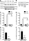
FAK inhibition abolishes VTN mediated LIF and Il-6 induction. C6 cells were seeded onto VTN-coated tissue culture plates (50 µg/ml) or non-treated control (Ctrl) plates and maintained for 4 h. (A) In these conditions, VTN stimulates phosphorylation of Y397-FAK (pFAK) but not of PYK2 at the corresponding Y402 residue (pPYK2), as shown in western blots. Loading controls are total FAK (tFAK) and PYK2 (tPYK2). Two representative lanes per group from three independent experiments are shown (n=3). (B) FAK antagonist PF573228 (10 µM, FAKi) completely blocked the VTN-stimulated pY397-FAK formation when added immediately after cells were seeded (n=3). In addition, the FAK inhibitor PF573228 (PF228) completely abolished VTN-induced LIF (C) and IL-6 (D) gene expression as measured by RT-qPCR. Control (Ctrl) vehicle is 0.1% DMSO (mean±s.e.m.; n=4 independent experiments). In further experiments, C6 cells were cultured in serum-free medium for 24 h before VTN was added directly to medium with or without FAK inhibition (PF228), (E) LIF and IL-6 (F) induction by VTN was also completely abolished by FAK inhibition in these conditions (mean±s.e.m.; n=3).
Fig. 5.
Fig. 5.
FAK mediates injury-induced LIF and IL-6 induction in C6 cells. C6 cells were seeded and maintained for 48 h in serum-containing medium without added VTN. Cells were then injured in an in vitro trauma model (swipe injury) with or without FAK inhibitors added at the time of injury. LIF (A) and IL-6 (B) mRNA expression were strongly induced (Ctrl Inj) at 4 h after injury compared to no injury controls (Ctrl NI), but were abolished by treatment with FAK antagonists, PND-1186 (PND), PF573228 (PF228), PF562271 (PF271), but not Y11. Surprisingly, Y11 further increased IL-6 expression after injury. Data are means±s.e.m. of three independent experiments and expressed as a fold change relative to uninjured controls, first normalized to GAPDH to account for differences in cell numbers. *P<0.05; **P<0.01; ***P<0.001; NS, not significant. In the same conditions as described for A and B, LIF (C) and IL-6 (D) protein expression and release was increased after injury, and attenuated by all inhibitors, except Y11, as shown by dot blots of conditioned medium (representative of three independent experiments). In addition, we found that C6 cells overexpressing wild-type (WT) FAK expressed significantly more LIF (E) and IL-6 (F) than cells overexpressing dominant negative Y397F mutant FAK following injury (mean±s.e.m.; n=3).
Fig. 6.
Fig. 6.
FAK, but not PYK2, regulates LIF and IL-6 expression in human and mouse brain endothelial cells. Human CMEC cells were passaged and resuspended in serum-free medium and maintained in suspension for 1 h. Cells were then plated onto plastic (PBS; Ctrl), or VTN-, laminin- (LN) or fibronectin (FN1)-treated plates for 1 h. (A) VTN caused a much greater increase in pFAK Y397 and STAT3 Y705 over laminin and fibronectin. CMEC cells were pre-incubated with integrin β3- or β5-blocking antibodies before plating onto VTN. At 4 h after plating, induction of LIF (B) and IL-6 (C) by VTN were reduced when CMEC cells were pre-incubated with P11, β3- or β5-blocking antibodies (mean±s.e.m.; n=4–6, ANOVA with Fisher LSD test). To determine the role and specifity of FAK in regulating baseline LIF and IL-6 mRNA expression, we performed knockdown of FAK (siFAK) or PYK2 (siPYK2) using siRNAs in human CMEC cells over 6 days. siFAK reduced LIF (A) and IL-6 (B), while increasing CNTF (C) gene expression relative to non-targeting control siRNA. siPYK2 had no effect. In bEnd5 cells, siFAK also diminished both LIF (D) and IL-6 (E) expression while increasing CNTF (F) (mean±s.e.m.; n=3). Pharmacological FAK inhibition for 4 h with PF573228 (PF228) also suppressed LIF (G) and IL-6 (H) while upregulating CNTF (I) mRNA expression relative to vehicle-treated controls (Ctrl), as measured by RT-qPCR (n=4). Data are means±s.e.m. from n=4 independent experiments each. *P<0.05, **P<0.01, ***P<0.001; NS, not significant.
Fig. 7.
Fig. 7.
Pharmacological FAK inhibitors are specific in suppressing LIF and IL-6 in CMEC cells. To confirm the specificity of PF573228 (PF228) and PND-1186 (PND), FAK was first knocked down in CMECs with siRNA for 5 days followed by 4 h inhibitor treatments. Total FAK (tFAK) and pFAK protein knockdown was quantified by capillary western blots (A). For illustration purposes, synthetic bands were produced from chemiluminescence spectrograms (B). TUB, α-tubulin. (C) Quantification showing that siFAK decreases total FAK normalized to α-tubulin, which is not affected by the inhibitors PF228 or PND. (D–F) Similarly, siFAK decreased pY397-FAK levels, with the PF228 and PND decreasing pFAK only under control siRNA conditions (siCtrl). Critically, LIF (G) and IL-6 (H) gene expression (RT-qPCR) were not significantly different when the inhibitors were incubated with siCtrl or siFAK, and provided no additive suppression in the presence of siFAK, showing that their effects were entirely mediated through FAK. Data are means±s.e.m. of four independent experiments and presented relative to DMSO- and control (siCtrl)-treated cells. *P<0.05; **P<0.01; NS, not significant.
Fig. 8.
Fig. 8.
Blood VTN leaks into the brain after stroke. (A) Schematic showing an overview of the signaling pathway by which VTN induces LIF and IL-6 gene expression, by binding to αvβ3 integrin and specifically activating downstream FAK. (B) Proposed model of VTN leakage after blood–brain barrier (BBB) breakdown. Under normal conditions, the intact BBB, characterized by tight junctions (TJ) between endothelial cells (EC), keeps VTN in blood from entering the central nervous system (CNS) tissue. BM, basement membrane. Under pathological conditions that cause BBB breakdown, such as stroke and hemorrhage, leakage of VTN in the brain parenchyma induces LIF and IL-6 expression by astrocytes (gray cells), microglia (blue cells) and endothelial cells (green cells).

Similar articles

Cited by

References

    1. Aguirre Ghiso J. A. (2002). Inhibition of FAK signaling activated by urokinase receptor induces dormancy in human carcinoma cells in vivo. Oncogene 21, 2513-2524. 10.1038/sj.onc.1205342 - DOI - PubMed
    1. Antonov A. S., Antonova G. N., Munn D. H., Mivechi N., Lucas R., Catravas J. D. and Verin A. D. (2011). αVβ3 integrin regulates macrophage inflammatory responses via PI3 kinase/Akt-dependent NF-κB activation. J. Cell. Physiol. 226, 469-476. 10.1002/jcp.22356 - DOI - PMC - PubMed
    1. Bang J.-Y., Kim E.-Y., Kang D.-K., Chang S.-I., Han M. H., Baek K.-H. and Kang I.-C. (2011). Pharmacoproteomic analysis of a novel cell-permeable peptide inhibitor of tumor-induced angiogenesis. Mol. Cell Proteomics 10, M110.005264 10.1074/mcp.M110.005264 - DOI - PMC - PubMed
    1. Banner L. R. and Patterson P. H. (1994). Major changes in the expression of the mRNAs for cholinergic differentiation factor/leukemia inhibitory factor and its receptor after injury to adult peripheral nerves and ganglia. Proc. Natl. Acad. Sci. USA 91, 7109-7113. 10.1073/pnas.91.15.7109 - DOI - PMC - PubMed
    1. Banner L. R., Moayeri N. N. and Patterson P. H. (1997). Leukemia inhibitory factor is expressed in astrocytes following cortical brain injury. Exp. Neurol. 147, 1-9. 10.1006/exnr.1997.6536 - DOI - PubMed

Publication types

MeSH terms