. 2017:82:223-234.
doi: 10.1101/sqb.2017.82.034132. Epub 2017 Dec 8.

Regulation of Crossover Frequency and Distribution during Meiotic Recombination

Affiliations

Regulation of Crossover Frequency and Distribution during Meiotic Recombination

Takamune T Saito et al. Cold Spring Harb Symp Quant Biol. 2017.

Abstract

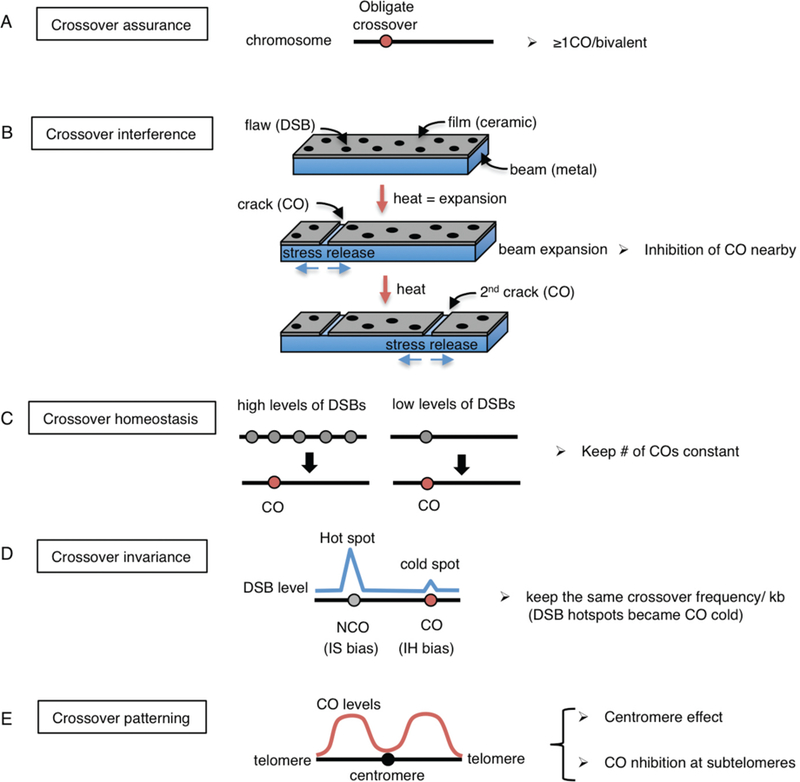
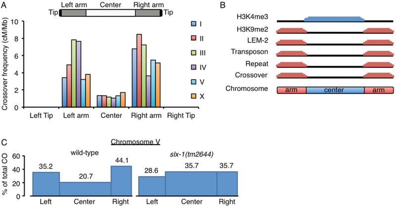
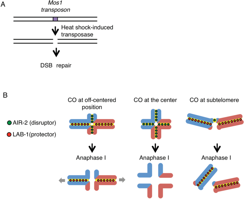
Crossover recombination is essential for generating genetic diversity and promoting accurate chromosome segregation during meiosis. The process of crossover recombination is tightly regulated and is initiated by the formation of programmed meiotic DNA double-strand breaks (DSBs). The number of DSBs is around 10-fold higher than the number of crossovers in most species, because only a limited number of DSBs are repaired as crossovers during meiosis. Moreover, crossovers are not randomly distributed. Most crossovers are located on chromosomal arm regions and both centromeres and telomeres are usually devoid of crossovers. Either loss or mislocalization of crossovers frequently results in chromosome nondisjunction and subsequent aneuploidy, leading to infertility, miscarriages, and birth defects such as Down syndrome. Here, we will review aspects of crossover regulation observed in most species and then focus on crossover regulation in the nematode Caenorhabditis elegans in which both the frequency and distribution of crossovers are tightly controlled. In this system, only a single crossover is formed, usually at an off-centered position, between each pair of homologous chromosomes. We have identified C. elegans mutants with deregulated crossover distribution, and we are analyzing crossover control by using an inducible single DSB system with which a single crossover can be produced at specific genomic positions. These combined studies are revealing novel insights into how crossover position is linked to accurate chromosome segregation.


Figures

Figure 1
Figure 1. Meiosis and crossover formation
Chromosome dynamics during meiosis. After premeiotic DNA replication, homologous chromosomes find each other (pairing) during the leptotene-zygotene stages. The synaptonemal complex assembles, aligning and holding homologs together throughout their full lengths (synapsis) at the pachytene stage. Repair of DNA double-strand breaks (DSBs) via crossover formation involves the reciprocal exchange of genetic information between homologs. A chiasma is the cytologically visible manifestation of an earlier crossover event underpinned by flanking sister chromatid cohesion and is observed as a cruciform configuration during the diplotene to diakinesis transition. Homologous chromosomes are segregated at the metaphase I to anaphase I transition and sister chromatids are separated at the metaphase II to anaphase II transition. Paternal chromatids are blue and maternal chromatids are red. Sister chromatid cohesion is depicted in yellow and the synaptonemal complex is depicted in green.
Figure 2
Figure 2. Model of homologous recombination.
DNA double strand breaks are generated by the topoisomerase-like protein Spo11. The MRN/X complex (Mre11-Rad50-Nbs1/Xrs2) resects the 5’ends to expose 3’overhangs. Single end invasion (SEI) is mediated by Rad51. Homologous recombination can then proceed through the following pathways: (A) synthesis-dependent strand annealing resulting in non-crossover products or (B) double Holliday junction (dHJ) formation by Mer3 and Msh4-Msh5 resulting in crossover (CO) formation. The DNA helicase ReqQ homologs Sgs1 and RTEL-1 disrupt D-loops to anneal both ends of the DSB. Once double Holliday junctions are formed, they are resolved by the structure-specific endonucleases SLX-1-SLX-4/HIM-18, MUS-81-EME1 and XPF-ERCC1. (C) Asymmetric resolution of the dHJ produces crossovers and (D) symmetric resolution results in non-crossovers. (E) dHJs can also be processed by the dissolution pathway through the BTR complex (BLM-TOP3-RMI1/2) to make non-crossover products. Paternal DNAs are blue and maternal DNAs are red. Circles indicate 5’side of DNA. Orange triangles indicate the direction of catalytic activities of Holliday junction resolvases. Key proteins acting at each step are indicated on the right and both yeast and worm names are indicated.
Figure 3
Figure 3. Different types of crossover control
Five known forms of crossover control are depicted. (A) Crossover assurance. At least one crossover per homologous chromosome pair is essential for chiasma formation and proper chromosome segregation at meiosis I. (B) Crossover interference. The beam-film model (modified from Kleckner et al. 2004) is represented. Chromosome axes and chromatin loops are likened as metallic beams and ceramic films which are tightly bonded to the beam, respectively. Heating the beam results in a flaw (DSB) being converted into a crack (CO formation), and the release of stress then propagates in both directions. Continued heating generates a 2nd crack away from the 1st crack resembling interference. (C) Crossover homeostasis. Either high or low levels of DSBs per homologous chromosome pair result in the same number of crossovers. Gray circles are DSBs and red circles are crossovers. (D) Crossover invariance. DSBs at hot spots tend to undergo intersister bias, resulting in a non-crossover outcome, while DSBs at cold spots undergo interhomolog bias leading to crossover products in S. pombe. (E) Crossover distribution/centromere effect. Crossovers near centromeres and telomeres are suppressed. Crossovers are also suppressed at the center regions in the holocentric organism C. elegans.
Figure 4
Figure 4. Tight regulation of crossover formation in C. elegans
(A) Crossovers are enriched at arm regions but suppressed at center regions in both autosomes and the X chromosome in C. elegans. No crossovers are observed at subtelomeric regions (average <614kb from telomeres). Data was adapted from (Rockman and Kruglyak 2009). (B) Unique features divide chromosome domains in C. elegans. Although up to ~10 DSBs are distributed in a non-biased manner along chromosomes (Saito et al. 2012), crossovers occur at the arm regions where the heterochromatin marker histone H3K9me2, the nuclear membrane protein (LEM-2) binding sequences, transposons, and repeat sequences, are enriched. Crossover formation is suppressed at the center region where the euchromatic marker histone H3K4me3 is enriched. (C) Crossover suppression at the center region of autosomes is lost in slx-1(tm2644) null mutants. Blue boxes indicate crossover frequencies (Saito et al. 2012). While the overall crossover frequency is not altered, crossover distribution is altered by increasing at the center region and decreasing at the arms in slx-1 mutants compared to wild type.
Figure 5
Figure 5. Site-specific analysis of meiotic recombination in C. elegans
(A) Mos1-based single inducible DSB system. Mos1 transposons and transposases are integrated into chromosomes in a spo-11 mutant background. The Mos1 integrated strain library is available at Nemagentag (http://elegans.imbb.forth.gr/nemagenetag/). Heat shock induces expression of the transposase which excises the Mos1 transposon resulting in a single DSB at a specific genomic position. (B) The single DSB system can be used to investigate the positional effect of crossovers on meiotic chromosome segregation. Blue and red lines are paternal and maternal chromatids, respectively. The system allows us to analyze the outcome of a single crossover forming at specific chromosomal sites. A crossover at the very center region disrupts the asymmetric configuration of the bivalent resulting in premature sister chromatid separation or homolog nondisjunction. Crossovers at subtelomeres result either in potentially fragile connections that are not stably retained at the ends of very short arms or events that fail to mature into crossovers at those positions.

References

    1. Acquaviva L, Szekvolgyi L, Dichtl B, Dichtl BS, de La Roche Saint Andre C, Nicolas A, Geli V 2013. The COMPASS subunit Spp1 links histone methylation to initiation of meiotic recombination. Science 339: 215–218. - PubMed
    1. Agostinho A, Meier B, Sonneville R, Jagut M, Woglar A, Blow J, Jantsch V, Gartner A. 2013. Combinatorial regulation of meiotic holliday junction resolution in C. elegans by HIM-6 (BLM) helicase, SLX-4, and the SLX-1, MUS-81 and XPF-1 nucleases. PLoS Genet 9: e1003591. - PMC - PubMed
    1. Barnes TM, Kohara Y, Coulson A, Hekimi S. 1995. Meiotic recombination, noncoding DNA and genomic organization in Caenorhabditis elegans. Genetics 141: 159–179. - PMC - PubMed
    1. Baudat F, Buard J, Grey C, Fledel-Alon A, Ober C, Przeworski M, Coop G, de Massy B. 2010. PRDM9 is a major determinant of meiotic recombination hotspots in humans and mice. Science 327: 836–840. - PMC - PubMed
    1. Beadle GW. 1932. A Possible Influence of the Spindle Fibre on Crossing-Over in Drosophila. Proc Natl Acad Sci U S A 18: 160–165. - PMC - PubMed

LinkOut - more resources