

DNA Methylation-Mediated Silencing of Regenerating Protein 1 Alpha (REG1A) Affects Gastric Cancer Prognosis
- PMID: 29222406
- PMCID: PMC5737223
- DOI: 10.12659/msm.904706
DNA Methylation-Mediated Silencing of Regenerating Protein 1 Alpha (REG1A) Affects Gastric Cancer Prognosis
Abstract
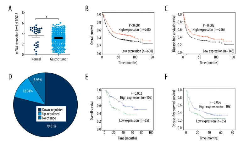
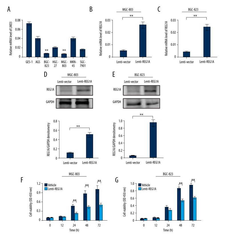
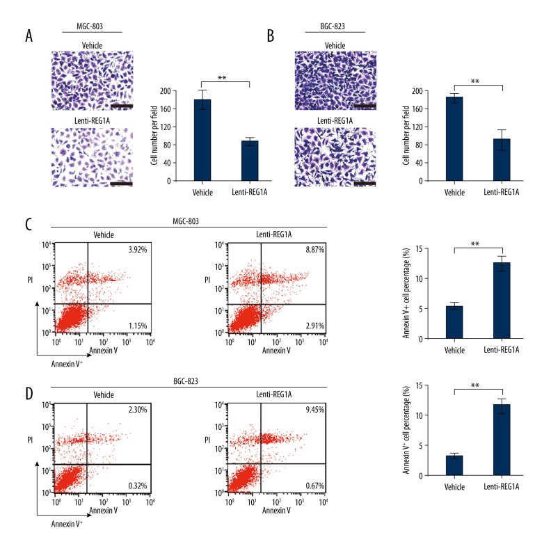
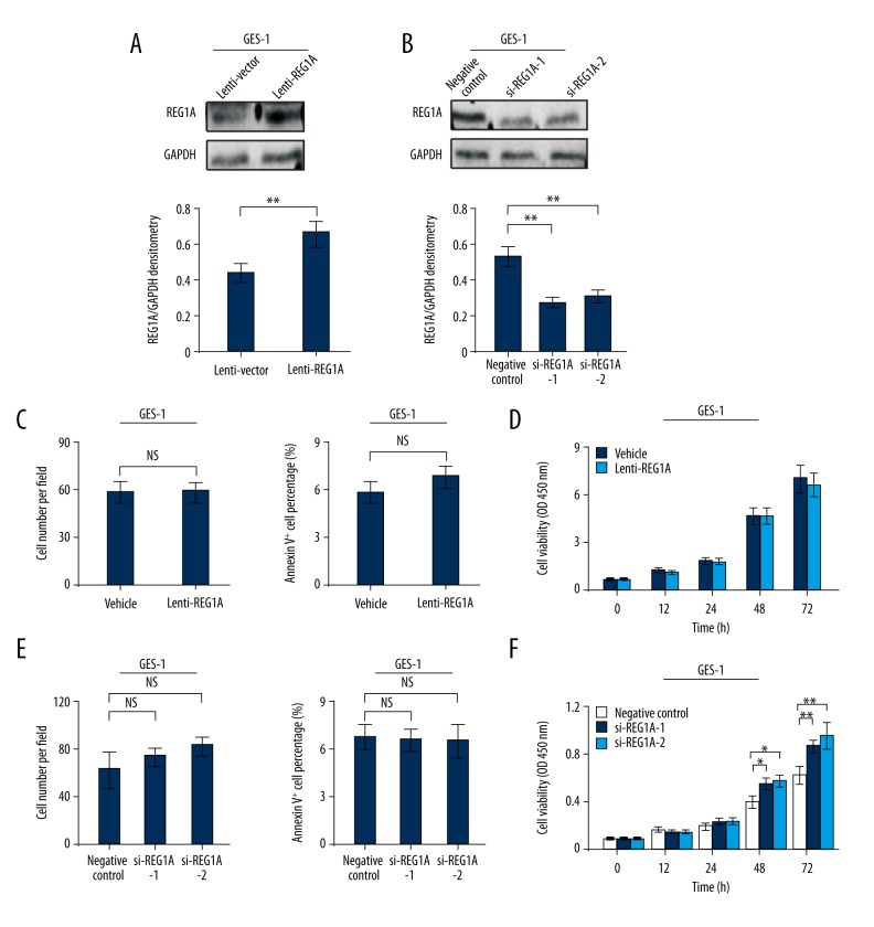
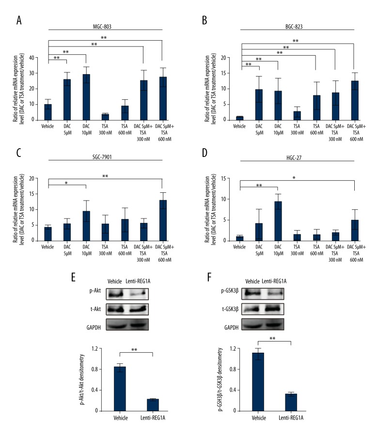
BACKGROUND Gastric cancer (GC) is one of the most common cause of cancer-related deaths. The clinical trials still lack the effective methods to treat or monitor the disease progression. In this research, the biological function and the underlying molecular mechanism of regenerating protein 1 alpha (REG1A) in GC were investigated. MATERIAL AND METHODS Gene expression omnibus (GEO), KMplot datasets and GC tissue microarray (n=164) were used to analyze the expression of REG1A and related patient prognoses in GC. Transwell matrigel assay, flow cytometry analysis and CCK8 cell viability assay were performed to detect the biological functions of REG1A. Western blotting and real-time PCR were used to detect the REG1A expression and PI3K/Akt related signaling. RESULTS It was found that the expression of REG1A was significantly downregulated in GC and closely related with clinicopathological findings or patient prognoses. REG1A overexpression could suppress the invasion, cell viability and promote the apoptosis of GC cells. Moreover, we found that the epigenetic methylation suppressed the expression level of REG1A in GC, and REG1A overexpression could suppress the phosphorylation of Akt or GSK3β signaling. CONCLUSIONS Taken together, REG1A regulates cell invasion, apoptosis and viability in GC through activating PI3K/Akt-GSK3β signaling. REG1A may serve as a promising therapeutic strategy for GC.
Conflict of interest statement
None.
Figures
Similar articles
-
KRAB zinc-finger protein 382 regulates epithelial-mesenchymal transition and functions as a tumor suppressor, but is silenced by CpG methylation in gastric cancer.Int J Oncol. 2018 Sep;53(3):961-972. doi: 10.3892/ijo.2018.4446. Epub 2018 Jun 19. Int J Oncol. 2018. PMID: 29956735 Free PMC article.
-
Epigenetic regulation of REG1A and chemosensitivity of cutaneous melanoma.Epigenetics. 2013 Oct;8(10):1043-52. doi: 10.4161/epi.25810. Epub 2013 Jul 31. Epigenetics. 2013. PMID: 23903855 Free PMC article.
-
MicroRNA-497 acts as a tumor suppressor in gastric cancer and is downregulated by DNA methylation.Oncol Rep. 2017 Jul;38(1):497-505. doi: 10.3892/or.2017.5698. Epub 2017 Jun 6. Oncol Rep. 2017. PMID: 28586056
-
lncRNA BG981369 Inhibits Cell Proliferation, Migration, and Invasion, and Promotes Cell Apoptosis by SRY-Related High-Mobility Group Box 4 (SOX4) Signaling Pathway in Human Gastric Cancer.Med Sci Monit. 2018 Feb 5;24:718-726. doi: 10.12659/msm.905965. Med Sci Monit. 2018. PMID: 29398692 Free PMC article.
-
DNA Methylation: An Important Biomarker and Therapeutic Target for Gastric Cancer.Front Genet. 2022 Mar 4;13:823905. doi: 10.3389/fgene.2022.823905. eCollection 2022. Front Genet. 2022. PMID: 35309131 Free PMC article. Review.
Cited by
-
Multiplex profiling of peritoneal metastases from gastric adenocarcinoma identified novel targets and molecular subtypes that predict treatment response.Gut. 2020 Jan;69(1):18-31. doi: 10.1136/gutjnl-2018-318070. Epub 2019 Jun 6. Gut. 2020. PMID: 31171626 Free PMC article.
-
Whole genome transcriptome analysis of the stomach resected in human vertical sleeve gastrectomy: cutting more than calories.Physiol Genomics. 2021 May 1;53(5):193-205. doi: 10.1152/physiolgenomics.00082.2020. Epub 2021 Apr 19. Physiol Genomics. 2021. PMID: 33870723 Free PMC article.
-
Four Decades After the Discovery of Regenerating Islet-Derived (Reg) Proteins: Current Understanding and Challenges.Front Cell Dev Biol. 2019 Oct 22;7:235. doi: 10.3389/fcell.2019.00235. eCollection 2019. Front Cell Dev Biol. 2019. PMID: 31696115 Free PMC article. Review.
-
E-cadherin variants associated with oral facial clefts trigger aberrant cell motility in a REG1A-dependent manner.Cell Commun Signal. 2024 Feb 27;22(1):152. doi: 10.1186/s12964-024-01532-x. Cell Commun Signal. 2024. PMID: 38414029 Free PMC article.
-
Matrine Alleviates Atherosclerosis by Targeting REG1A and Activating the PI3K/AKT/mTOR Pathway to Inhibit Endothelial Cell Ferroptosis.Biochem Genet. 2025 Apr 25. doi: 10.1007/s10528-025-11117-z. Online ahead of print. Biochem Genet. 2025. PMID: 40281246
References
-
- Wong SS, Kim KM, Ting JC, et al. Genomic landscape and genetic heterogeneity in gastric adenocarcinoma revealed by whole-genome sequencing. Nat Commun. 2014;5:5477. - PubMed
-
- Zang ZJ, Cutcutache I, Poon SL, et al. Exome sequencing of gastric adenocarcinoma identifies recurrent somatic mutations in cell adhesion and chromatin remodeling genes. Nat Genet. 2012;44(5):570–74. - PubMed
MeSH terms
Substances
LinkOut - more resources
Full Text Sources
Medical
Research Materials
Miscellaneous
