. 2017 Dec 8;8(1):1991.
doi: 10.1038/s41467-017-02223-7.

Immune stealth-driven O2 serotype prevalence and potential for therapeutic antibodies against multidrug resistant Klebsiella pneumoniae

Affiliations

Immune stealth-driven O2 serotype prevalence and potential for therapeutic antibodies against multidrug resistant Klebsiella pneumoniae

Meghan E Pennini et al. Nat Commun. .

Abstract

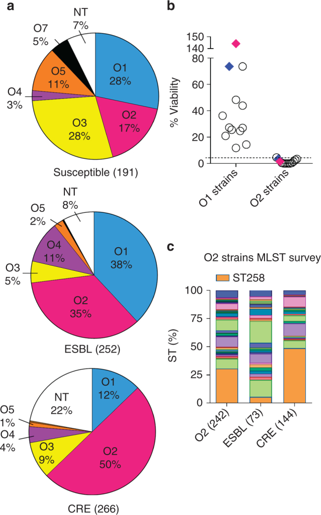
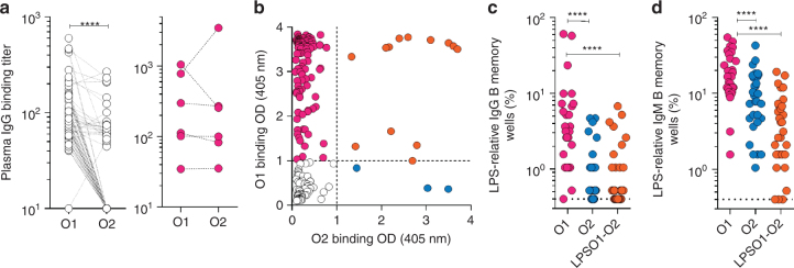
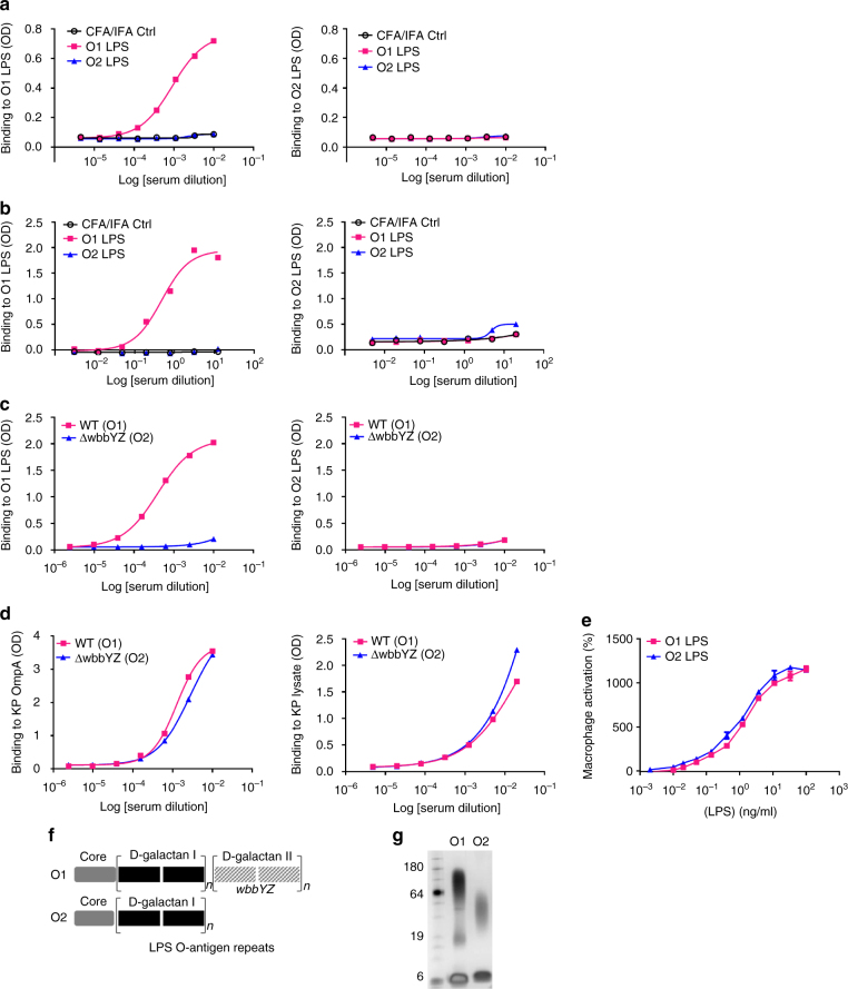
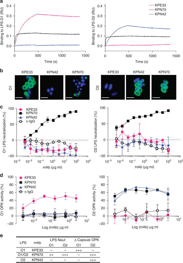
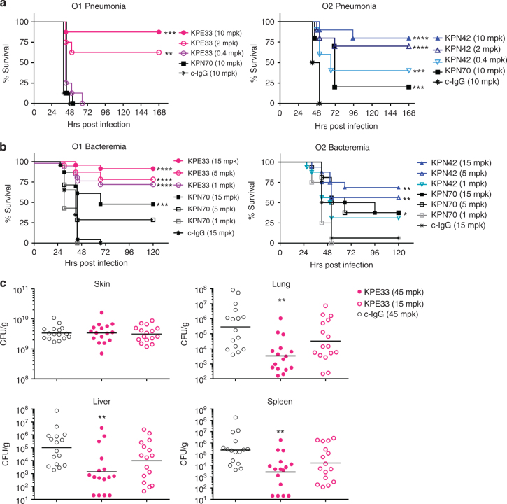
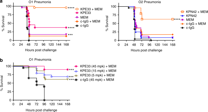
Emerging multidrug-resistant bacteria are a challenge for modern medicine, but how these pathogens are so successful is not fully understood. Robust antibacterial vaccines have prevented and reduced resistance suggesting a pivotal role for immunity in deterring antibiotic resistance. Here, we show the increased prevalence of Klebsiella pneumoniae lipopolysaccharide O2 serotype strains in all major drug resistance groups correlating with a paucity of anti-O2 antibodies in human B cell repertoires. We identify human monoclonal antibodies to O-antigens that are highly protective in mouse models of infection, even against heavily encapsulated strains. These antibodies, including a rare anti-O2 specific antibody, synergistically protect against drug-resistant strains in adjunctive therapy with meropenem, a standard-of-care antibiotic, confirming the importance of immune assistance in antibiotic therapy. These findings support an antibody-based immunotherapeutic strategy even for highly resistant K. pneumoniae infections, and underscore the effect humoral immunity has on evolving drug resistance.


Conflict of interest statement

M.E.P., Q.W., M.P.,J.B, R.C, E.S, X.X, W.Z., M.M.C., A.D., P.W., J.S., C.K.S. are current or former employees of MedImmune/AstraZeneca and may own stock or stock options in the company. Patents describing the activity of the antibodies in this work have been filed by MedImmune. A.D.M., M.B, E.C., S.B, F.Z, A.L, D.C. are employees of HumAbs BioMed and may currently hold Humabs stocks or stock options. The remaining authors declare no competing financial interests.

Figures

Fig. 1
Fig. 1
Klebsiella pneumoniae serotype O2 prevalence in MDR strains. a LPS serotype-specific antibodies were used to determine the serotype of 709 K. pneumoniae clinical isolates from 38 different countries by western blot. Pie charts represent the distribution of each serotype as a percentage of the total in each antibiotic resistant group. Susceptible, ESBL and CRE strains are categorized based on their MIC profile as described in Methods. More information about the source of the isolates is listed in Supplementary Table 1. b Serum sensitivity assays were performed in 45% pooled human sera using a panel of 12 O1 isolates, 12 O2 isolates and two separate isogenic pairs genetically converting an O2 strain to O1 (Kp961842, magenta diamonds) or an O1 strain to O2 (Kp1131115, blue diamonds). Percent viability is the ratio between CFUs present in buffer control media and CFUs present in human sera after 2 h growth. c A subset of serotype O2 isolates was further analyzed by Sanger or next generation sequencing to determine ST types. The CRE ST258 sequence type is represented by the shaded orange portion of each bar to highlight the percent of ST258 isolates observed in each category
Fig. 2
Fig. 2
Analysis of the human antibody response to K. pneumoniae O1 and O2 LPS. a O1 and O2-specific IgG antibodies were measured by ELISA in plasma derived from 103 healthy donors (left panel) and 6 ICU patients (right panel) diagnosed with O2 K. pneumoniae infection. Shown are the endpoint titers based on serial dilutions. Lines between data points indicate data acquired from the same sample. bd Analysis of antigen-specific repertoires (AMBRA, antigen-specific memory B cell repertoire analysis) from tonsillar IgG and IgM memory B cells of 33 donors (30 of the 33 were available for IgM analysis, Supplementary Fig. 2). Total tonsillar lymphocytes were plated and stimulated with R848 and IL-2 and the 10-day culture supernatants (192 cultures per donor) were analyzed by ELISA for the presence of IgG or IgM antibodies that bind to O1 LPS, O2 LPS or uncoated control plates. The OD values obtained from the analysis of uncoated plates were subtracted from the analysis of the O1 and O2 specific responses. b Shown are ELISA OD values of individual cultures from the analysis of one representative donor. Cut-off values are indicated by a dotted line. O1, O2 and O1/O2 reactive cultures are represented as magenta, blue and orange circles, respectively. c, d Each data point represents the frequency of the cultures (n = 192) scoring as positive for IgG (c) or IgM (d) reactivity to O1 or O2 LPS for each of the donors analyzed, according to the cut-off values shown in b. Data was analyzed using the Wilcoxon matched-pair signed rank test to assess significance. ****p < 0.0001
Fig. 3
Fig. 3
K. pneumoniae O2 LPS is less immunogenic than K. pneumoniae O1 LPS. Purified O1 and O2 LPS were used for immunization of mice (a) or rabbits (b) with 0.5 mg LPS per animal. Immune sera were collected from O1 and O2 immunized animals and used to assess antibody titer against KP O1 or O2 LPS by ELISA. Sera from mock immunized animals with adjuvant only (black open circle) were used as a negative control. c Mice were immunized subcutaneously with either heat killed Kp1131115 (WT, an O1 strain) or the isogenic matched Kp1131115ΔwbbYZ (ΔwbbYZ, O2 serotype). Sera were collected 7 days after the final injection and used to assess antibody titer against K. pneumoniae O1 or O2 LPS by ELISA. d Sera collected in c were used to assess antibody binding to a protein target (OmpA) and to proteinase K bacterial lysate by ELISA. e Immune activation of cells in vitro was compared for purified O1 and O2 LPS. RAW264.7 cells stably transfected with a NF-κB driven luciferase promoter were stimulated with various doses of K. pneumoniae O1 or K. pneumoniae O2 LPS for 2 h. Percent activation was calculated as the amount of luminescence in stimulated cells vs. non-stimulated. Error bars indicate s.d. of each data point. f A schematic representation of KP O1 and O2 LPS. The gray region represents the membrane proximal LPS core region, the black region represents the d-galactan I (d-gal I) repeating O antigen sugars, the hatched gray filled region represents the O1 specific d-galactan II (d-gal II) repeating O antigen sugars encoded by the wbbYZ locus. Deletion of this locus converted the O1 strain (Kp1131115 WT) to an O2 expressing strain (Kp1131115ΔwbbYZ). g Silver stain of a SDS-PAGE gel showing the relative molecular weight of K. pneumoniae O1 LPS (lane 2) vs. K. pneumoniae O2 LPS (lane 3)
Fig. 4
Fig. 4
In vitro characterization of anti-O antigen mAbs. a Association/dissociation curves of K. pneumoniae O1 or K. pneumoniae O2 LPS binding to mAbs KPE33, KPN70, and KPN42 were measured by ForteBio Octet using protein A capture probe pre-loaded with each mAb. b Confocal images of mAb binding to an O1 (Kp43816) or O2 (Kp9148) strain (scale bar, 2 μm). c LPS neutralization was measured using RAW264.7 cells transfected with a NK-kB driven luciferase promoter stimulated with 2 ng ml−1 of purified O1 or O2 LPS in the presence of KPE33, KPN70, KPN42 or the isotype-matched control mAb (c-IgG). Percent inhibition was calculated as the amount of luminescence (RLU) in treated wells divided by the RLU in wells stimulated with LPS alone multiplied by 100. d Opsonophagocytic killing assays were performed using various concentrations of mAbs in the presence of the phagocytic cell line HL-60. The capsule-deficient luminescent mutants Kp43816ΔcpsB and Kp8570ΔcpsB were used as the O1 and O2 serotype targets, respectively. Percent killing was calculated based on the amount of luminescence after 2.5 h incubation in treated wells vs. wells with bacteria only. An isotype-matched mAb (c-IgG) is used as a negative control. e Summary of the in vitro functional activities against O1 and O2 K. pneumoniae for each mAb. All error bars indicate s.d. at each data point
Fig. 5
Fig. 5
Anti-O1 and anti-O2 mAbs are protective in vivo. a Therapeutic activity of the mAbs was tested in C57BL/6 mice infected intranasally with a K. pneumoniae O1 (Kp1131115, 6 × 107 CFU) or O2 (Kp961842, 2 × 108 CFU) strain. KPE33 (anti-O1), KPN42 (anti-O2) and KPN70 (anti-O1/O2) mAbs were administered intravascular 1 h post infection at the doses indicated. An isotype-matched mAb (c-IgG) was used as a negative control. b Prophylactic activity of the mAbs was tested in C57BL/6 mice infected intraperitoneal with the O1 strain Kp1131115 (3 × 106 CFU) or the O2 strain Kp961842 (1 × 107 CFU). mAbs were administered IP 24 h prior to infection at the doses indicated; survival was monitored for 5 days post infection. c CF1 mice received a dorsal burn followed by K. pneumoniae O1 (Kp1131115, 5 × 106) bacterial inoculation subcutaneously at the burn site. KPE33 (45 or 15 mpk) or isotype-matched control mAb (c-IgG, 45 mpk) were administered IP 24 h post infection, and organs were harvested 48 h post infection to determine bacterial CFU. All graphs are representative of at least three separate experiments. Mantel-Cox analysis was done to assess significant survival benefit compared to the control mAb. For thermal injury CFU comparison, Kruskal Wallace one way Anova analysis was done to compare differences in CFU between KPE33 treated vs. control IgG treated groups, ****p < 0.0001, ***p < 0.001, **p < 0.01
Fig. 6
Fig. 6
Antibody synergy with antibiotic. a Mice were infected intranasally as in Fig. 5 with the K. pneumoniae O1 (Kp1131115, 6 × 107 CFU) or O2 (Kp961842, 2 × 108 CFU) strain then treated 1 h post infection with suboptimal doses of mAb (KPE33, 1 mg kg−1 or KPN42, 0.2 mg kg−1 given IV) and antibiotic (meropenem, 7.5 mg kg−1 for O1 strain; 50 mg kg−1 for O2 strain, given subcutaneous) or a combination of both mAb and antibiotic. Graphs represent the combined data from three separate experiments with a total of 24 mice in each group. Mantel-Cox analysis was done to assess significant survival benefit compared to the control mAb (a) or control mAb + MEM (b), ****p < 0.0001, ***p < 0.001, **p < 0.01. The Bliss independence method was used to determine synergistic activity of the combination therapy vs. either monotherapy alone; p = 2.5 × 10−6 for KPE33+MEM combination and p = 7.7 × 10−5 for KPN42+MEM combination. b Mice were infected intransal with a KP O1 strain (Kp8045, 1 × 104 CFU). Meropenem (1.5 mpk) was administered subcutaneous 4 h post infection and mAb KPE33 was administered IV 24 h post infection at the indicated doses. All graphs are representative of at least three separate experiments. An isotype-matched mAb (c-IgG) was used as a negative control. Mantel-Cox analysis was done to assess significant survival benefit compared to the control mAb+MEM b, ****p < 0.0001, ***p < 0.001, **p < 0.01

References

    1. McKenna M. Antibiotic resistance: the last resort. Nature. 2013;499:394–396. doi: 10.1038/499394a. - DOI - PubMed
    1. Iredell J, Brown J, Tagg K. Antibiotic resistance in Enterobacteriaceae: mechanisms and clinical implications. BMJ. 2016;352:h6420. doi: 10.1136/bmj.h6420. - DOI - PubMed
    1. Taur Y, Pamer EG. The intestinal microbiota and susceptibility to infection in immunocompromised patients. Curr. Opin. Infect. Dis. 2013;26:332–337. doi: 10.1097/QCO.0b013e3283630dd3. - DOI - PMC - PubMed
    1. Badal RE, et al. Etiology, extended-spectrum beta-lactamase rates and antimicrobial susceptibility of Gram-negative bacilli causing intra-abdominal infections in patients in general pediatric and pediatric intensive care units--global data from the Study for Monitoring Antimicrobial Resistance Trends 2008 to 2010. Pediatr. Infect. Dis. J. 2013;32:636–640. doi: 10.1097/INF.0b013e3182886377. - DOI - PubMed
    1. van Duin D, Paterson DL. Multidrug-resistant bacteria in the community: trends and lessons learned. Infect. Dis. Clin. North Am. 2016;30:377–390. doi: 10.1016/j.idc.2016.02.004. - DOI - PMC - PubMed

Publication types

MeSH terms