Review
. 2018 Feb 1;140(2):0208031-02080313.
doi: 10.1115/1.4038704.

Elastic Fibers and Large Artery Mechanics in Animal Models of Development and Disease

Affiliations
Review

Elastic Fibers and Large Artery Mechanics in Animal Models of Development and Disease

Maria Gabriela Espinosa et al. J Biomech Eng. .

Abstract

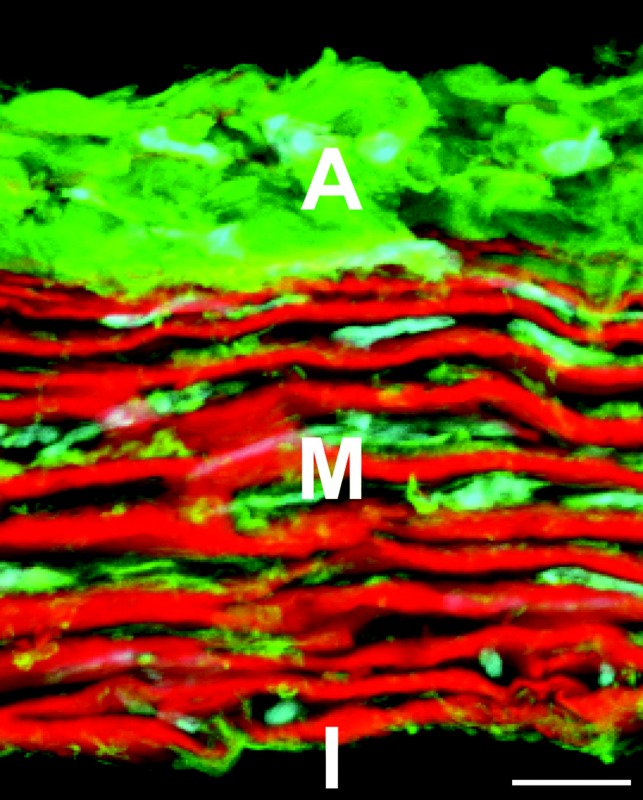
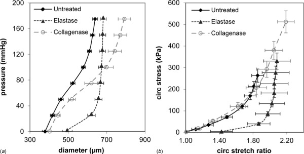
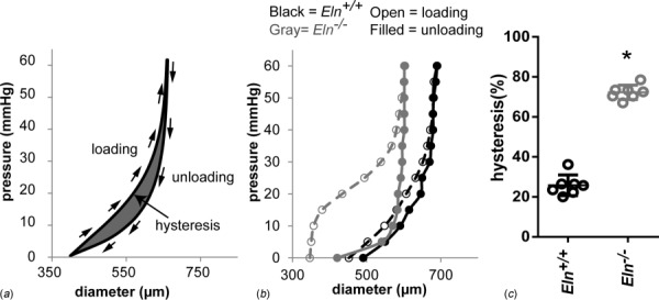
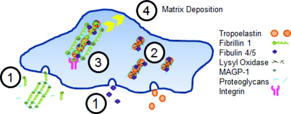
Development of a closed circulatory system requires that large arteries adapt to the mechanical demands of high, pulsatile pressure. Elastin and collagen uniquely address these design criteria in the low and high stress regimes, resulting in a nonlinear mechanical response. Elastin is the core component of elastic fibers, which provide the artery wall with energy storage and recoil. The integrity of the elastic fiber network is affected by component insufficiency or disorganization, leading to an array of vascular pathologies and compromised mechanical behavior. In this review, we discuss how elastic fibers are formed and how they adapt in development and disease. We discuss elastic fiber contributions to arterial mechanical behavior and remodeling. We primarily present data from mouse models with elastic fiber deficiencies, but suggest that alternate small animal models may have unique experimental advantages and the potential to provide new insights. Advanced ultrastructural and biomechanical data are constantly being used to update computational models of arterial mechanics. We discuss the progression from early phenomenological models to microstructurally motivated strain energy functions for both collagen and elastic fiber networks. Although many current models individually account for arterial adaptation, complex geometries, and fluid-solid interactions (FSIs), future models will need to include an even greater number of factors and interactions in the complex system. Among these factors, we identify the need to revisit the role of time dependence and axial growth and remodeling in large artery mechanics, especially in cardiovascular diseases that affect the mechanical integrity of the elastic fibers.


Figures

Fig. 1
Fig. 1
Cross section of the mouse ascending aorta stained with fluorescent probes for elastin (red), collagen (green), and cell nuclei (cyan). Adventitia (A), media (M), and intima (I) are shown. The image is a maximum projection of several z-planes for a slice that is not a perfect circumferential cross section, which provides the illusion of depth. Scale bar = 10 μm.
Fig. 2
Fig. 2
Mechanical contributions of elastin and collagen in the mouse carotid artery. Diameter–pressure (a) and circumferential stretch ratio–Cauchy stress (b) relationships are shown. Arteries were untreated or treated with elastase or collagenase to digest elastin or collagen, respectively, and then mounted in a pressure myograph. Elastase treated arteries dilate at low pressures and then become very stiff at high pressures, while collagenase treated arteries show little change at low pressures and dilate at high pressures, becoming less stiff. The results are consistent with the idea that elastin and collagen dominate the circumferential mechanical behavior at low and high pressures, respectively. N = 6–8/group. Mean ± SEM.
Fig. 3
Fig. 3
Hysteresis is increased in the absence of elastin. Example loading and unloading curves showing the hysteresis, or area between the loading and unloading diameter–pressure curves (a). Representative diameter–pressure curves for loading and unloading of a newborn aorta from Eln+/+ and Eln−/− mice show the increased hysteresis area in Eln−/− aorta (b). Quantification of the hysteresis area as a percent of the total area under the loading curve shows a three-fold increase in hysteresis for Eln−/− aorta compared to Eln+/+ (c). Hysteresis is indicative of the energy loss during cyclic loading. N = 6–7/group. * = P <0.05 by students t-test [34].
Fig. 4
Fig. 4
Model of elastic fiber assembly. Microfibrils consisting mostly of fibrillin-1, but also including MAGP-1 and interacting with proteoglycans, are assembled in the extracellular space. At the same time, fibulin-4/5 and tropoelastin are secreted from the SMC (1). Tropoelastin coacervates and then interacts with fibulin-4/5 and is crosslinked by LOX on the SMC surface (2). Integrins link microfibrils to the cell surface where they interact with the tropoelastin/fibulin aggregates and allow further crosslinking by LOX (3). The crosslinked, fully assembled elastic fiber is then deposited into the ECM (4).
Fig. 5
Fig. 5
Microfibrillar organization in developing chick DA. Chick embryos at embryonic day 6 (a) and 10 (b) were sectioned at the level of the heart and the DA was stained with fluorescently tagged antibodies to cell nuclei (blue) and fibrillin-2 (purple). The density and circumferential orientation of the fibrillin-2 microfibrillar scaffold increase over the four day developmental period. The DA lumen is at the bottom of the images. Scale bar = 10 μm.
Fig. 6
Fig. 6
Configurations for arterial growth and remodeling. Each constituent, elastin (E), muscle (SMCs) (m), and collagen (C) has its own stress-free configuration (denoted by capital B). For muscle, BP is the passive stress-free configuration and BA is the active stress-free configuration. Stressed configurations (denoted by lower case b's) vary with time and are color coded. Blue denotes configuration at time <0, green at time = 0, yellow at time = 0 ≤ τt, and red at time = t. Muscle undergoes several different stressed configurations including bν = unloaded and intact, bR = unloaded after growth and activation that induces residual stress, and bL = loaded. Stretch ratios between states are denoted by λ's with superscripts denoting the constituent and subscripts denoting the time or mode of deformation: g = growth, a = activation, A = active, and P = passive. λ* for each constituent is the stretch ratio from the stress-free configuration to the loaded configuration of the composite arterial wall. From Alford et al. [144]. Reprinted with permission from Springer @ 2008.

References

    1. Yurchenco, P. D. , and O'Rear, J. J. , 1994, “ Basal Lamina Assembly,” Curr. Opin. Cell Biol., 6(5), pp. 674–681.10.1016/0955-0674(94)90093-0 - DOI - PubMed
    1. Han, S. , Shin, Y. , Jeong, H. E. , Jeon, J. S. , Kamm, R. D. , Huh, D. , Sohn, L. L. , and Chung, S. , 2015, “ Constructive Remodeling of a Synthetic Endothelial Extracellular Matrix,” Sci. Rep., 5, p. 18290.10.1038/srep18290 - DOI - PMC - PubMed
    1. Schwartz, S. M. , and Benditt, E. P. , 1972, “ Studies on Aortic Intima—I: Structure and Permeability of Rat Thoracic Aortic Intima,” Am. J. Pathol., 66(2), pp. 241–264.https://www.ncbi.nlm.nih.gov/pubmed/5009972 - PMC - PubMed
    1. Gerrity, R. G. , Richardson, M. , Somer, J. B. , Bell, F. P. , and Schwartz, C. J. , 1977, “ Endothelial Cell Morphology in Areas of In Vivo Evans Blue Uptake in the Aorta of Young Pigs—II: Ultrastructure of the Intima in Areas of Differing Permeability to Proteins,” Am. J. Pathol., 89(2), pp. 313–334.https://www.ncbi.nlm.nih.gov/pmc/articles/PMC2032231/ - PMC - PubMed
    1. Levesque, M. J. , and Nerem, R. M. , 1985, “ The Elongation and Orientation of Cultured Endothelial Cells in Response to Shear Stress,” ASME J. Biomech. Eng., 107(4), pp. 341–347.10.1115/1.3138567 - DOI - PubMed

Publication types

LinkOut - more resources