Longevity of visual improvements following transcorneal electrical stimulation and efficacy of retreatment in three individuals with retinitis pigmentosa
- PMID: 29222719
- PMCID: PMC6039224
- DOI: 10.1007/s00417-017-3858-8
Longevity of visual improvements following transcorneal electrical stimulation and efficacy of retreatment in three individuals with retinitis pigmentosa
Abstract
Purpose: A small-scale randomized controlled trial conducted by our group found that four of seven retinitis pigmentosa (RP) subjects who received six weekly Transcorneal Electrical Stimulation (TES) sessions developed significant improvements in visual acuity (VA), quick contrast sensitivity function (qCSF), and/or Goldmann visual fields (GVF). We longitudinally monitored three of these participants for declining visual function due to natural RP progression to determine the duration of their responses and administered retreatments.
Methods: Over a period of 29-35 months, repeated ETDRS VA, qCSF and/or GVF tests and three to six TES treatment courses consisting of six weekly sessions were administered in each eye of three RP participants every four to 16 months in an unmasked, prospective case series study.
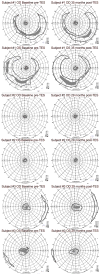
Results: For two participants, there were significant VA improvements of 44-52 letters (0.88-1.04 logMAR) and 15-23 letters (0.3-0.46 logMAR) in the worse eye at baseline after each of three or four treatment courses of TES compared to initial baseline. They had no significant decreases from baseline for VA or qCSF over 29 to 35 months, The third participant had a significant mean improvement in VA in the eye with better baseline vision (p = 0.004) and binocularly (p < 0.001) following six treatment courses over the 29-month period. For the first two participants, mean annual rates of GVF change for each eye ranged from -5% to 0% with the V4e stimulus, and -26% to +33% the III4e stimulus. The third participant's mean annual GVF changes were +14 to +35%, with a statistically significant improvement across 29 months for both the V4e and III4e stimuli in the right eye (p = 0.045; p = 0.015) and the V4e stimulus in the left eye (p = 0.047).
Conclusion: Following encouraging visual improvements after TES that lasted for several months, it appears it may be possible to restore and prevent slowly diminishing vision over time with retreatments, which requires confirmation in a large-scale randomized controlled trial.
Keywords: Contrast sensitivity; Goldmann visual field; Retinitis pigmentosa; Transcorneal electrical stimulation; Visual acuity.
Figures


References
-
- Tao Y, Chen T, Liu ZY, et al. Topographic quantification of the Transcorneal electrical stimulation (TES)-induced protective effects on N-methyl-N-Nitrosourea-treated retinas. Invest Ophthalmol Vis Sci. 2016;57(11):4614–4624. - PubMed
-
- Schatz A, Röck T, Naycheva L, et al. Transcorneal electrical stimulation for patients with retinitis pigmentosa: a prospective, randomized, sham-controlled exploratory study. Invest Ophthalmol Vis Sci. 2011;52(7):4485–4496. - PubMed
-
- Schatz A, Pach J, Gosheva M, et al. Transcorneal electrical stimulation for patients with retinitis Pigmentosa: a prospective, randomized, sham-controlled follow-up study over 1 year. Invest Ophthalmol Vis Sci. 2017;58(1):257–269. - PubMed
-
- Robles-Camarillo D, Niño-de-Rivera L, López-Miranda J, et al. The effect of transcorneal electrical stimulation in visual acuity: retinitis pigmentosa. J Biomed Sci Eng. 2013;6:1–7.
Publication types
MeSH terms
Grants and funding
LinkOut - more resources
Full Text Sources
Other Literature Sources
Medical

