. 2018:1727:179-200.
doi: 10.1007/978-1-4939-7571-6_14.

Diolistic Labeling and Analysis of Dendritic Spines

Affiliations

Diolistic Labeling and Analysis of Dendritic Spines

M Foster Olive et al. Methods Mol Biol. 2018.

Abstract

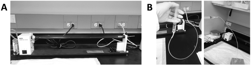
Dendritic spines are diverse and plastic components of the neuronal cell apparatus and are highly responsive to trophic factors during both development and adulthood. Diolistic labeling of neurons with lipophilic fluorescent dyes, coupled with advanced high-resolution microscopy methods, provides robust labeling of dendritic spines for assessment of their density and morphology. Here, we describe a method for labeling of dendritic spines using diolistic labeling in ex vivo brain slices, visualization using confocal laser scanning microscopy, deconvolution, and analysis using the Surpass module of Bitplane Imaris software.

Keywords: 3-D reconstruction; Confocal imaging; Deconvolution; Dendritic spine; Diolistic; Lipophilic dye; Morphology.


Figures

Figure 1.
Figure 1.
(A) Tubing preparation station (BioRad). (B) Filling of Tefzel tubing with PVP solution via withdrawing it from the microcentrifuge tube with a syringe.
Figure 2.
Figure 2.
(A) Use of a razor blade to finely chop tungsten particles on a glass microscope slide. (B) Appearance of tungsten particles after addition of dichloromethane.
Figure 3.
Figure 3.
(A) Placement of coated tubing into tubing cutter (BioRad). (B) Appearance of bullets, which should be stored in a light-proof container with a dessicant at room temperature.
Figure 4.
Figure 4.
Sectioning of 1.5% PFA-fixed brain tissue on a vibratome. Coronal brain sections through the dorsal and ventral striatum can be observed at the bottom of the photograph, one of which overlying the bristles of a paintbrush which is used to manipulate the sections.
Figure 5.
Figure 5.
BioRad Helios gene gun with modified barrel and attached nylon mesh filter.
Figure 6.
Figure 6.
Proper placement of gene gun barrel with filter tip for labeling of tissue section.
Figure 7.
Figure 7.
Cropping of the acquired dendrite image in the xy (upper arrow) and z (lower arrow) axes.
Figure 8.
Figure 8.
Example of image parameters in Summary tab of Data Manager window in AutoQuant.
Figure 9.
Figure 9.
Selection of Surpass mode and option for using the mouse pointer in Select mode (for individual points on dendrites) or Navigate mode (for 3-D rotation of image).
Figure 10.
Figure 10.
Add New Filament icon for creation of selected dendritic segment.
Figure 11.
Figure 11.
Manual selection of dendritic segment, selection of Autopath mode of detection, and assignment of selection as a dendrite.
Figure 12.
Figure 12.
Selection of start and end points of dendritic segments for analysis.
Figure 13.
Figure 13.
Assignment of selected starting point as a segment.
Figure 14.
Figure 14.
Centering the line within the selected dendritic segment.
Figure 15.
Figure 15.
Style and Edit menus for filament selection.
Figure 16.
Figure 16.
Process selection menu for selection of spine diameter settings.
Figure 17.
Figure 17.
Spine diameter settings (minimum, maximum, and threshold).
Figure 18.
Figure 18.
Correct and incorrect filling of dendritic shaft with the analysis line.
Figure 19.
Figure 19.
Style menu for initiating spine analysis using Autodepth method.
Figure 20.
Figure 20.
A. Selection of the terminal point of an individual dendritic spine. B. Filling of individual spine (blue color). C. Filling of all spines.
Figure 21.
Figure 21.
A. Inversion of dendritic selection color from yellow to gray. B. Selection of all dendritic spines as indicated by a change from blue to yellow fill color.
Figure 22.
Figure 22.
Appropriately filled dendritic spines. Not the variation in thickness of spine heads, necks, and dendrite attachment points.
Figure 23.
Figure 23.
A. Selection (A) and assignment (B) of dendrite segment end point. C. Marked dendritic segment endpoint.
Figure 24.
Figure 24.
Selection of filament data set for export.
Figure 25.
Figure 25.
A. Selection of data analysis tool. B. Exporting all dendritic spine data to Excel.

References

    1. Cho C, MacDonald R, Shang J, Cho MJ, Chalifour LE, Paudel HK (2017) Early growth response-1-mediated down-regulation of drebrin correlates with loss of dendritic spines. J Neurochem, in press. - PubMed
    1. Andero R, Choi DC, Ressler KJ (2014) BDNF-TrkB receptor regulation of distributed adult neural plasticity, memory formation, and psychiatric disorders. Prog Mol Biol Transl Sci 122:169–192. - PubMed
    1. Gomez-Palacio-Schjetnan A, Escobar ML (2013) Neurotrophins and synaptic plasticity. Curr Top Behav Neurosci 15:117–136. - PubMed
    1. Harward SC, Hedrick NG, Hall CE, Parra-Bueno P, Milner TA, Pan E, Laviv T, Hempstead BL, Yasuda R, McNamara JO (2016) Autocrine BDNF-TrkB signalling within a single dendritic spine. Nature 538 (7623):99–103. - PMC - PubMed
    1. Irala D, Bonafina A, Fontanet PA, Alsina FC, Paratcha G, Ledda F (2016) The GDNF-GFRalpha1 complex promotes the development of hippocampal dendritic arbors and spines via NCAM. Development 143 (22):4224–4235. - PubMed

Publication types

Substances

LinkOut - more resources