. 2018 Nov;28(6):791-805.
doi: 10.1111/bpa.12583. Epub 2017 Dec 28.

Dominant role of microglial and macrophage innate immune responses in human ischemic infarcts

Affiliations

Dominant role of microglial and macrophage innate immune responses in human ischemic infarcts

Tobias Zrzavy et al. Brain Pathol. 2018 Nov.

Abstract

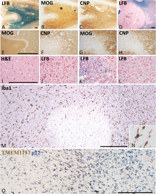
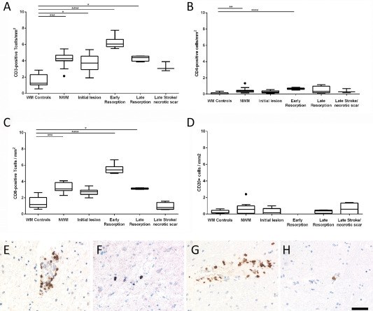
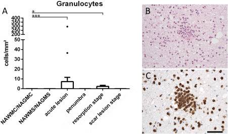
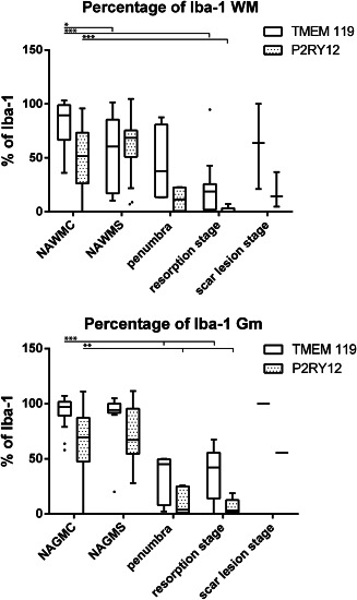
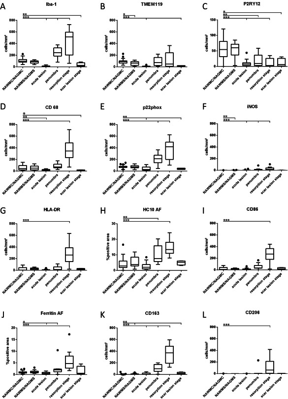
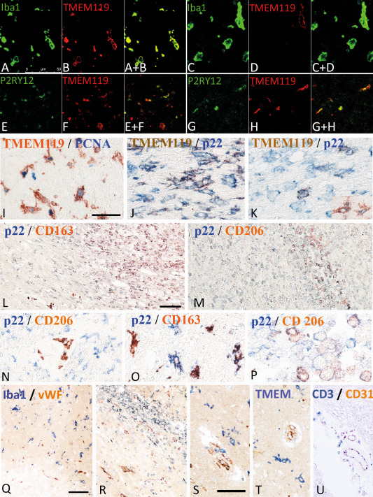
Inflammatory mechanisms, involving granulocytes, T-cells, B-cells, macrophages and activated microglia, have been suggested to play a pathogenic role in experimental models of stroke and may be targets for therapeutic intervention. However, knowledge on the inflammatory response in human stroke lesions is limited. Here, we performed a quantitative study on the inflammatory reaction in human ischemic infarct lesions. We found increased numbers of T-lymphocytes, mainly CD8+ cells, but not of B-lymphocytes. Their number was very low in comparison to that seen in inflammatory diseases of the central nervous system and they did not show signs of activation. Polymorphonuclear leukocytes were present in meninges and less prominently in the perivascular space in early lesions, but their infiltration into the lesioned tissue was sparse with the exception of a single case. Microglia were lost in the necrotic core of fresh lesions, their number was increased in the surrounding penumbra, apparently due to proliferation. Using TMEM119 as a marker for the resident microglia pool, macrophages in lesions were in part derived from the original microglia pool, depending on the lesion stage. Most microglia and macrophages revealed a pro-inflammatory activation pattern, expressing molecules involved in phagocytosis, oxidative injury, antigen presentation and iron metabolism and had partially lost the expression of P2RY12, an antigen expressed on homeostatic ("resting") microglia in rodents. At later lesion stages, the majority of macrophages showed intermediate activation patterns, expressing pro-inflammatory and anti-inflammatory markers. Microglia in the normal white matter of controls and stroke patients were already partly activated toward a pro-inflammatory phenotype. Our data suggest that the direct contribution of lymphocytes and granulocytes to active tissue injury in human ischemic infarct lesions is limited and that stroke therapy that targets pro-inflammatory microglia and macrophage activation may be effective.

Keywords: P2RY12; Stroke; TMEM119; granulocytes; lymphocytes; macrophages; microglia.


Conflict of interest statement

The authors have no conflict of interest.

Figures

Figure 1
Figure 1
Neuropathological basis of the staging of acute ischemic infarct lesions. A–C. 85 year old femal patient with a stroke history of 31 days. Parietal cortex and subcortical white matter with multiple acute and subscute ischemic infarct lesions due to thrombosis of meningeal artery. Multiple focal lesions in the cortex and the subcortical white matter. Some of the lesions are in the initial stage of tissue necrosis, defined by reduced myelin staining in luxol fast blue and loss of CNPase expression, while MOG is still preserved (*); More advanced lesions show complete loss of myelin and a parallel loss of CNP and MOG (+). D. 85 year old female with a stroke history of 8 days; acute cortico‐subcortical ischemic infacr lesion with some hemorrhagic component in the cortical lesion part. E,F. Higher magnification of the lesion indicated by * in Figure 1A; the acute ischemic lesion is demarcated from the surrounding normal tissue by a band of tissue edema; within the lesion there is a complete loss of CNPase, but a preservation of MOG immunoreactivity; G,H. higher magnification of the lesion indicated by + in Figure 1; there is a complete loss of both myelin proteins (MOG in g) and CNP in h); the lesion appears hypercellular due to the macrophage infiltration present in the resorption stage; A–H. Magnification bar: 1 mm. I. Acute ischemic infarct lesion in the cortex of the patient, shown in Figure 1D; profound reduction of cell density in the cortex and in particular a profound loss of neurons and pale neurons with nuclear fragmentation and some apoptotic nuclei of glia cells; hematoxlyin & eosin staining; J–L. Different lesions in the resorption stage in a 97 year old female patient; (J) Cortical lesion characterized by a high density of activated microglia and macrophages, some of them containing early and late lipid degradation products; (K) White matter lesion in the early resorption stages with macrophages, containing luxol fast blue reactive degradation products; (L) White matter lesion in the late resorption stages with macrophages containing empty lipid vacuoles; M,N. Microglia reaction within and around the acute ischemic inflarct lesion, shown by * in Figure 1A; the lesion is located at the border between white and grey matter; within the lesions Iba‐1 postive cells are completely lost; at the lesion edge microglia starts to increase in density and many of these cells are double labeled with the proliferation marker PCNA (N). O. Double staining for TMEM119 and NADPHoxidase (p22phox) in a lesion at the early resorption stage. Microglia is found in the perilesional area (left side). At the edge of the lesion, a profound increase of microglia and macrophage like cells is present, the majority of those still being double stained for both markers. In the center of the lesions, numerous macrophages are present, which are p22phox+/TMEM119 cells. (C–O) Magnification bar:200 μm.
Figure 2
Figure 2
T‐ and B‐Lymphocyte infiltration in the normal white and grey matter of controls and in human ischemic infarct lesions. A–D. Only few T‐ and B‐lymphocytes were present in the brain tissue of age matched controls and in different stages of ischemic infarct lesions, defined in material and methods. However, the number of infitrating T‐cells (cells/mm2) was significantly increased, reaching its peak at the stage of early resorption (A–C), while this was not the case for B‐cells (D). The graphs show CD3+ cells (a), CD8+ cells (B), CD4+ cells (C) and CD20+ B‐cells (D). The lower panels show examples for lymphocyte infiltration in the lesions; the sections were stained for CD3: E, CD4; F, CD8: G, and CD20: H. Magnification bar: 50 μm.
Figure 3
Figure 3
A. Tissue infiltration with granulocytes. In most of the cases, granulocytes were very sparse and this was the case during all lesion stages. Only a single case showed substantial numbers of granulocyte within the core of an acute lesion (single point with 295 cells/mm2). B,C. Show images of granulocyte infiltration in the case with substantial numbers of such cells, depicted in the staining with hematoxylin/eosin (B) and with p22phox (C). Magnification bar: 100 μm.
Figure 4
Figure 4
Percentage of Iba‐1+ cells expressing TMEM119 or P2RY12, in the brain tissue of controls and stroke patients and in different stages of ischemic infarct lesions. There is a significant reduction of the percentage of Iba‐1+ cells expressing these markers in the normal brain tissue of stroke and this percentage is further reduced within the lesions.
Figure 5
Figure 5
Quantitative profile of microglia/macrophage marker expression in the brain tissue of controls (WAWMC/NAWGS) and different stages of ischemic infarct lesions. For most markers, the values represent actual numbers of cells/mm2 in the different lesion areas. For the MHC Class I marker HC10 and for Ferritin (H,J), densitometry was performed, since these markers were also expressed on other resident cells of the brain. In these panels, the values represent the densitometric values, determined by area fraction (AF, see Material and Methods). *: P ≤ 0.05; **: P ≤ 0.01; ***: P ≤ 0.001.
Figure 6
Figure 6
Double staining of microglia and macrophages with different phenotypic markers. A–D. Double staining with the pan microglia/macrophage marker Iba‐1 and the marker for resident microglia derived cells, TMEM119. While in the normal brain tissue in this case around acute stroke lesions the majority of cells co‐express both markers (A,B), the number of macrophage like cells expressing TMEM119 is low in lesions in the late resorption stage (C,D). The double stained images are shown in A+B and c+d. E–H. Double staining for the “homeostatic” marker P2RY12 with TMEM119 shows that a fraction of microglia derived cells in the penumbra of acute lesions (E,F) and in the late scar stage (G,H) co‐express both markers. The double stained images are shown in E+F and G+H. I. Double staining for TMEM 119 and the proliferation marker PCNA. Many microglia in the penumbra around acute ischemic infarct lesions show a nuclear expression of PCNA. J,K. Higher magnification of lesion areas presented in Figure 1O, showing partial double staining with TMEM119 and p22phox in microglia like cells at the ledge of the lesions (J), while the number of double stained cells in the center of the lesions is low (K). L–P. Double staining for the pro‐inflammatory marker p22phox and the anti‐inflammatory markers CD206 (M, N, P) and CD163 (L,O) in ischemic infarct lesions, in the early and late resorption stage. While pro‐inflammatory markers are mainly expressed in the perilesional areas and in the early lesion stages, the anti‐inflammatory markers are mainly present in the center of the lesions, depicting the late resorption stage; l: low magnification of the lesion edge; M = low magnification of the lesion center; N = perilesional tissue with p22phox expression in activated microglia and CD206 expression in perivascular macrophages; O = lesion edge with microglia cells, some of them being double stained; Q = lesion center with macrophage like cells, some only expressing p22phox, while others also express CD206. Q–U. Double staining for T‐cells, macrophages and microglia with the vascular markers von Willebrand factor and CD31: (Q) Acute ischemic infarct lesion with loss of microglia in the ischemic core (lower corner of the image) and acitvated microglia in the penumbra; there is no preferential accumulation of myeloid cells around the vessels; (R) Ischemic infarct lesion in the eraly resorption stage; there are few perivascular myeloid cells, but no preferential accumlation of these cells around smaller vessels. S–U. Perivascular versus parenchymal location of Iba‐1+ myeloid cells (s), TMEM119+ microglia (T) and CD3+ T‐cells (U); Iba‐1+ cells are seen in perivascular and parenchymal location, while TMEM 119+ cells are only present in the parenchyma; CD3+ cells are in the vast majority located around medium sized veins of the tissue. All magnification bars represent 50 μm.

Similar articles

Cited by

References

    1. Aboul‐Enein F, Rauschka H, Kornek B, Stadelmann C, Stefferl A, Bruck W et al (2003) Preferential loss of myelin‐associated glycoprotein reflects hypoxia‐like white matter damage in stroke and inflammatory brain diseases. J Neuropathol Exp Neurol 62:25–33. - PubMed
    1. Bauer J, Lassmann H (2016) Neuropathological techniques to investigate central nervous system sections in multiple sclerosis. Methods Mol Biol 1304:211–229. - PubMed
    1. Bauer J, Stadelmann C, Bancher C, Jellinger K, Lassmann H (1999) Apoptosis of T lymphocytes in acute disseminated encephalomyelitis. Acta Neuropathol 97:543–546. - PubMed
    1. Bennett ML, Bennett FC, Liddelow SA, Ajami B, Zamanian JL, Fernhoff NB et al (2016) New tools for studying microglia in the mouse and human CNS. Proc Natl Acad Sci U S A 113:E1738–E1746. - PMC - PubMed
    1. Booss J, Esiri MM, Tourtellotte WW, Mason DY (1983) Immunohistological analysis of T lymphocyte subsets in the central nervous system in chronic progressive multiple sclerosis. J Neurol Sci 62:219–232. - PubMed

Publication types

MeSH terms