Review
. 2018 Mar:84:329-340.
doi: 10.1016/j.ultras.2017.11.013. Epub 2017 Nov 23.

Recent technological advancements in cardiac ultrasound imaging

Affiliations
Review

Recent technological advancements in cardiac ultrasound imaging

Jaydev K Dave et al. Ultrasonics. 2018 Mar.

Abstract

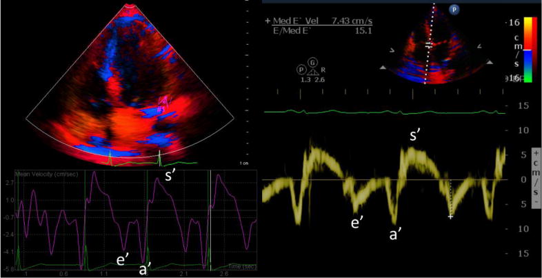
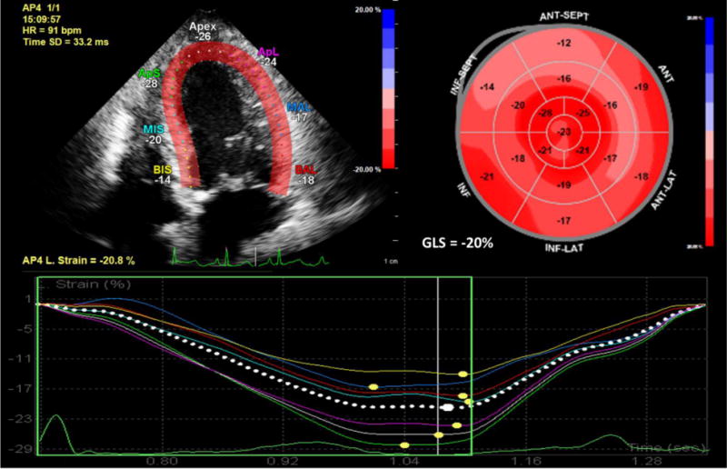
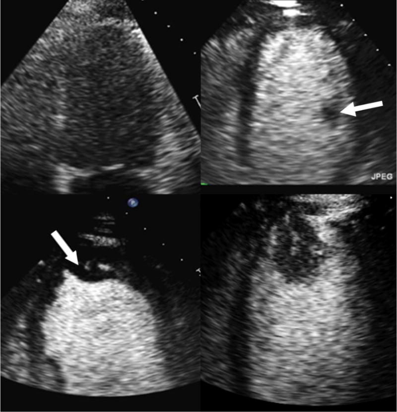
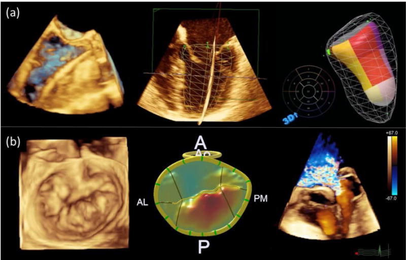
About 92.1 million Americans suffer from at least one type of cardiovascular disease. Worldwide, cardiovascular diseases are the number one cause of death (about 31% of all global deaths). Recent technological advancements in cardiac ultrasound imaging are expected to aid in the clinical diagnosis of many cardiovascular diseases. This article provides an overview of such recent technological advancements, specifically focusing on tissue Doppler imaging, strain imaging, contrast echocardiography, 3D echocardiography, point-of-care echocardiography, 3D volumetric flow assessments, and elastography. With these advancements ultrasound imaging is rapidly changing the domain of cardiac imaging. The advantages offered by ultrasound imaging include real-time imaging, imaging at patient bed-side, cost-effectiveness and ionizing-radiation-free imaging. Along with these advantages, the steps taken towards standardization of ultrasound based quantitative markers, reviewed here, will play a major role in addressing the healthcare burden associated with cardiovascular diseases.

Keywords: 3D echocardiography; 3D volumetric flow assessments; Cardiac elastography; Cardiac ultrasound; Contrast echocardiography; Point-of-care echocardiography; Strain imaging; Tissue Doppler imaging.


Figures

Figure 1
Figure 1
Color-coded tissue Doppler curves are displayed on the left after image post-processing. Pulsed-wave tissue Doppler curve is shown on the right with sample volume placed at the septal mitral annulus (e′=early diastolic velocity, a′=late diastolic velocity, and s′=systolic velocity; the peak after s′ and before e′ corresponds to the peak in the isovolumic relaxation phase, and the peak after a′ and before s′ corresponds to the peak in the isovolumic contraction phase).
Figure 2
Figure 2
Longitudinal strain by two-dimensional speckle-tracking echocardiography is shown in the apical four chamber view in top left. Bull’s eye map showing individual longitudinal strain values for all myocardial segments with color overlay and global longitudinal strain is shown in the top right. Longitudinal strain curves for ventricular segments in the four chamber view are shown at the bottom.
Figure 3
Figure 3
Endocardial borders are not well visualized without contrast (top left). Contrast microbubbles opacify the left ventricle improving delineation of endocardial borders (top right). A papillary muscle (white arrow), a normal intracardiac structure, is outlined by the contrast. Contrast enables clear visualization of an apical thrombus (arrow; bottom left) and apical tumor (bottom right). The presence of a vascular channel and enhancement with contrast help to differentiate apical tumor from apical thrombus.
Figure 4
Figure 4. 3D Echocardiographic Images
Panel a (top): Volume rendering showing wire in left ventricle during transcatheter aortic valve replacement (left); wire-frame surface rendering superimposed over multiplanar two-dimensional views of left ventricle (middle); and color-coded surface rendering of left ventricle with each color representing a single ventricular segment (right). Panel b (bottom): Volume rendering of mitral valve depicting posterior-leaflet mitral valve prolapse (left); surface rendering of mitral valve with color gradations representing degree of prolapse relative to annular plane (middle); and volume rendering with color Doppler depicting severe mitral regurgitation due to mitral valve prolapse (right).

References

    1. Hanna IR, Silverman ME. A history of cardiac auscultation and some of its contributors. Am J Cardiol. 2002;90:259–267. - PubMed
    1. Meyer RA. History of ultrasound in cardiology. J Ultrasound Med. 2004;23:1–11. - PubMed
    1. Roelandt JR. Seeing the invisible: a short history of cardiac ultrasound. Eur J Echocardiogr. 2000;1:8–11. - PubMed
    1. Mehta NJ, Khan IA. Cardiology’s 10 greatest discoveries of the 20th century. Tex Heart Inst J. 2002;29:164–171. - PMC - PubMed
    1. Picano E. Economic and biological costs of cardiac imaging. Cardiovasc Ultrasound. 2005;3:13. - PMC - PubMed