. 2018 Feb 2;8(2):567-575.
doi: 10.1534/g3.117.300325.

Growth of Caenorhabditis elegans in Defined Media Is Dependent on Presence of Particulate Matter

Affiliations

Growth of Caenorhabditis elegans in Defined Media Is Dependent on Presence of Particulate Matter

Matthew R Flavel et al. G3 (Bethesda). .

Abstract

Caenorhabditis elegans are typically cultured in a monoxenic medium consisting of live bacteria. However, this introduces a secondary organism to experiments, and restricts the manipulation of the nutritional environment. Due to the intricate link between genes and environment, greater control and understanding of nutritional factors are required to push the C. elegans field into new areas. For decades, attempts to develop a chemically defined, axenic medium as an alternative for culturing C. elegans have been made. However, the mechanism by which the filter feeder C. elegans obtains nutrients from these liquid media is not known. Using a fluorescence-activated cell sorting based approach, we demonstrate growth in all past axenic C. elegans media to be dependent on the presence of previously unknown particles. This particle requirement of C. elegans led to development of liposome-based, nanoparticle culturing that allows full control of nutrients delivered to C. elegans.

Keywords: Caenorhabditis elegans; axenic media; feeding; liposomes; nutrition.


Figures

Figure 1
Figure 1
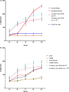
Effect of milk separation on growth rate: growth rate (length micrometer) of C. elegans measured at 24-hr intervals, while exposed to various dietary conditions. SD expressed as error bars. n = 25 per treatment group, growth in S– Medium containing OP50 E. coli (9 × 108 and 4.5 × 109 colonies per milliliter) included as positive control.
Figure 2
Figure 2
Effect of particulate matter on development: (A) Solubilization of particulate fraction in milk arrests C. Elegans growth. Growth rate (length micrometer) of C. elegans measured at 24 hr intervals, while exposed to CeHR media with added milk particulate matter that has either been solubilized with high concentration urea (8 M) and then diluted to 46 mM (solubilized media) or 46 mM urea added to CHR media + milk pellet, leaving milk pellet particles present (control media). SD expressed as error bars. n = 25 per treatment group. (B) Effect of filtration of AXM and CeMM media on C. elegans growth rate. Growth rate (length micrometer) of C. elegans measured at 24-hr intervals, while exposed to AXM media, CeMM media, AXM media filtered or CeMM filtered. SD expressed as error bars. n = 25 per treatment group, Growth in S– Medium containing OP50 E. coli (9 × 108 and 4.5 × 109 colonies per milliliter) included as positive controls in both figures.
Figure 3
Figure 3
FACS detection of particles in various media conditions. Distribution of different particle sizes. Purple, 1 μm; Green, 0.5 μm; Blue, 0.05 μm; Red, sample. (A) CeHR, (B) CeHR Filtered, (C) CeMM, (D) CeMM filtered, (E) CeMM Filtered and incubated at 20°, (F) = AXM, (G) AXM Filtered, (H) AXM Filtered and incubated at 20°. (I) Liposomes packed with CeHR. (J) Liposomes packed with M9.
Figure 4
Figure 4
Growth rate (length micrometer) of C. elegans measured at 24 hr while exposed to various media conditions. CeHR, AXM, and CeMM media were both filtered following the same protocol as used in Figure 1 and Figure 2B. However, each medium was then packed inside liposomes and suspended in M9 buffer. CeHR medium with milk and cholesterol removed were also packed within liposomes and suspended in M9 buffer. M9 Buffer or CeHR without liposomes were included as negative controls. n = 25 per treatment group, SD presented as error bars. Growth in S-medium containing OP50 E. coli (9 × 108 and 4.5 × 109 colonies per milliliter) in addition to CeHR prepared according to standard protocols included as positive controls.

References

    1. Avery L., 1993. Motor neuron M3 controls pharyngeal muscle relaxation timing in Caenorhabditis elegans. J. Exp. Biol. 175: 283–297. - PubMed
    1. Avery L., Shtonda B. B., 2003. Food transport in the C. elegans pharynx. J. Exp. Biol. 206: 2441–2457. - PMC - PubMed
    1. Avery, L., and Y. J. You, 2012 C. elegans feeding (May 21, 2012). WormBook, ed. The C. elegans Research Community WormBook, /10.1895/wormbook.1.150.1, http://www.wormbook.org.
    1. Baugh L. R., 2013. To grow or not to grow: nutritional control of development during Caenorhabditis elegans L1 arrest. Genetics 194: 539–555. - PMC - PubMed
    1. Brans G., Schroën C. G. P. H., Van der Sman R. G. M., Boom R. M., 2004. Membrane fractionation of milk: state of the art and challenges. J. Membr. Sci. 243: 263–272.

Publication types

MeSH terms