. 2017 Dec 20;96(6):1282-1289.e4.
doi: 10.1016/j.neuron.2017.11.006. Epub 2017 Dec 7.

Injecting Instructions into Premotor Cortex

Affiliations

Injecting Instructions into Premotor Cortex

Kevin A Mazurek et al. Neuron. .

Abstract

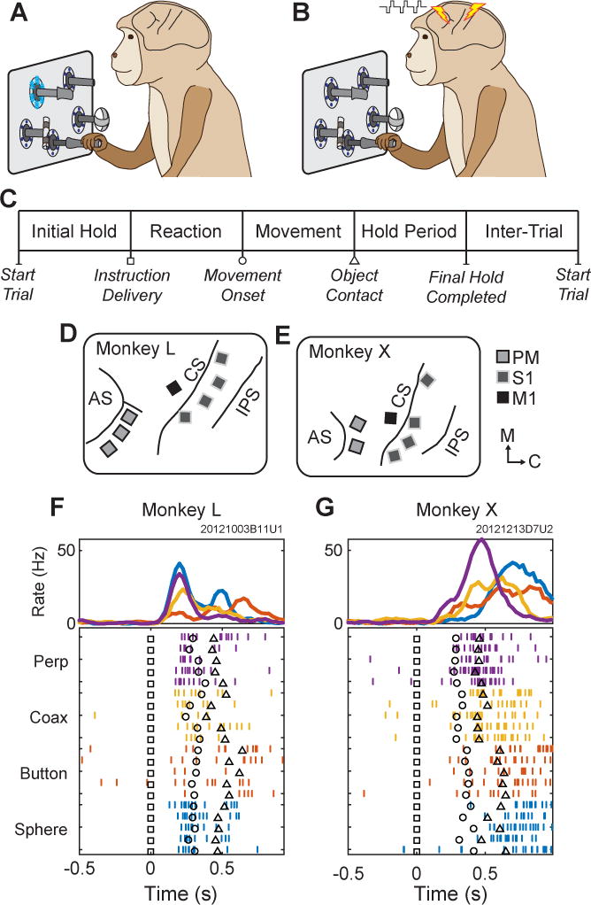
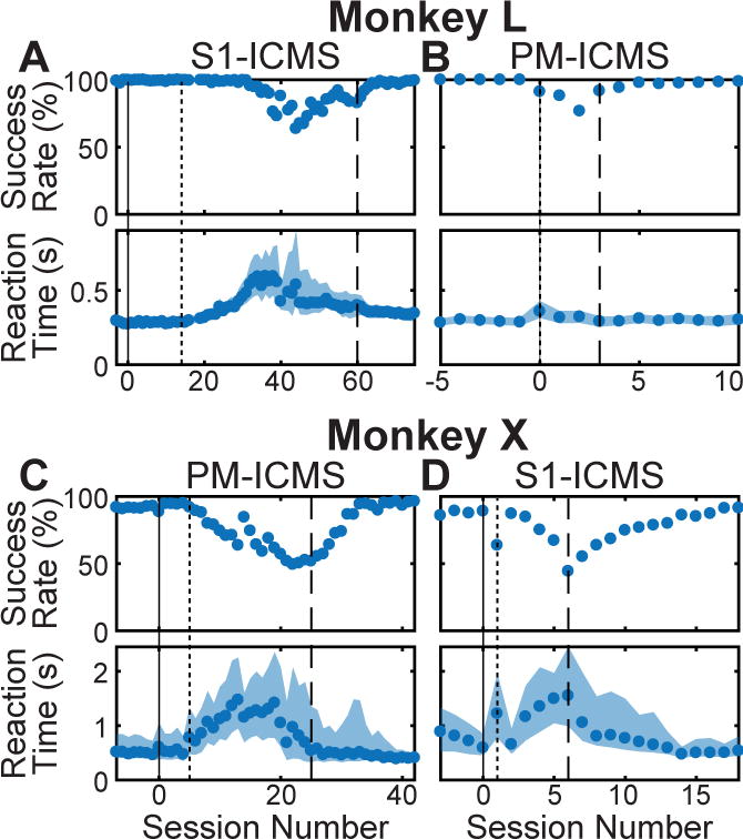
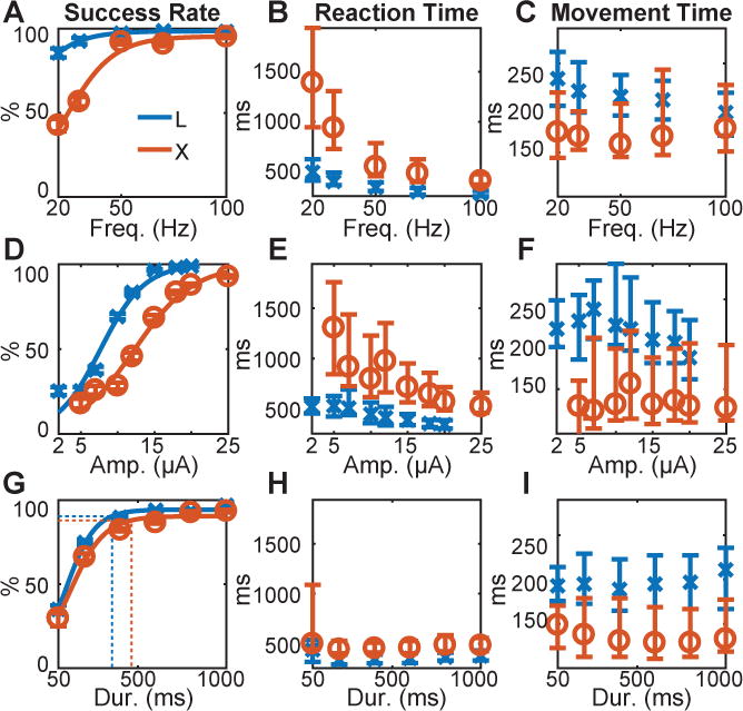
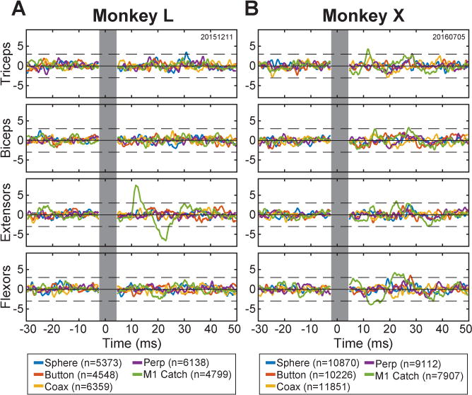
The premotor cortex (PM) receives inputs from parietal cortical areas representing processed visuospatial information, translates that information into programs for particular movements, and communicates those programs to the primary motor cortex (M1) for execution. Consistent with this general function, intracortical microstimulation (ICMS) in the PM of sufficient frequency, amplitude, and duration has been shown to evoke complex movements of the arm and hand that vary systematically depending on the locus of stimulation. Using frequencies and amplitudes too low to evoke muscle activity, however, we found that ICMS in the PM can provide instructions to perform specific reach, grasp, and manipulate movements. These instructed actions were not fixed but rather were learned through associations between the arbitrary stimulation locations and particular movements. Low-amplitude ICMS at different PM locations thus evokes distinguishable experiences that can become associated with specific movements arbitrarily, providing a novel means of injecting information into the nervous system.

Keywords: conditional association; grasping; intracortical microstimulation; learning; manipulation; premotor cortex; primary motor cortex; primary somatosensory cortex; reaching.


Figures

Figure 1
Figure 1
Task, instructions, and electrode locations. (A) Monkeys initially performed the reach-grasp-manipulate task instructed by blue LEDs. (B) Monkeys then learned to perform the same task instructed by ICMS delivered in either PM or S1. (C) Sequence of task epochs. (D,E) Floating microelectrode array locations in (D) monkey L and (E) monkey X. Light gray squares with dark outlines represent arrays in PM; dark gray squares with light outlines, S1, and black squares, M1. AS-arcuate sulcus, CS-central sulcus, IPS-intraparietal sulcus. Orientation arrows: M–medial, C–caudal. (F,G) Typical unit activity recorded from a PM electrode in each monkey during RGM movements (LED Instructions) involving each object: perpendicular cylinder (Perp, purple); coaxial cylinder (Coax, yellow); button (red); and sphere (blue). Only five trials of each movement are shown in the raster display, whereas ~20 trials of each are averaged in the histograms. Trials have been aligned at the instruction onset (Time=0, black squares in the raster trials). Additional markers indicate the time of movement onset (black circle) and target object contact (black triangle) in each trial.
Figure 2
Figure 2
Learning to use PM- or S1-ICMS instructions. In early sessions, only LED instructions were delivered. Beginning at session 0 (solid vertical line), both LED and ICMS instructions were delivered concurrently. Instruction LEDs then were gradually dimmed (beginning at dotted vertical line) and eventually were turned off completely (dashed vertical line). Monkey L first learned to use (A) S1-ICMS then (B) PM-ICMS. Monkey X first learned to use (C) PM-ICMS then (D) S1-ICMS. Reaction times are displayed as medians. The shaded regions represent the 25th to 75th percentile. See also Tables S1 and S1.
Figure 3
Figure 3
Parameter sweeps. Three parameters of PM-ICMS instructions were varied for each monkey: (A,B,C) pulse frequency, (D,E,F) current amplitude, and (G,H,I) train duration. Success rate (A,D,G), reaction time (B,E,H), and movement time (C,F,I), each have been plotted as a function of the swept parameter. In (G) the vertical dotted lines represent the median reaction time across all swept durations for each monkey (H), and the corresponding horizontal dotted lines indicate the success rate at this median reaction time, based on the fitted logistic functions. X’s (monkey L, blue) and O’s (monkey X, red) indicate medians; error bars represent the 25th and 75th percentiles. See also Table S3.
Figure 4
Figure 4
Stimulus-triggered averages of EMG activity. Averages of rectified EMG activity from four upper extremity muscle groups—Triceps, Biceps, forearm Extensors, and forearm Flexors—were compiled for each monkey (A, monkey L; B, monkey X) using individual ICMS pulses as triggers. ICMS parameters used for these sessions are given in Table S4. Separate averages (colors) were compiled using n pulses delivered through the PM electrode instructing the sphere, button, coaxial cylinder, or perpendicular cylinder, as well as for catch-trial pulses delivered in M1. The vertical gray bar indicates the interval during which the aligned stimulation artifacts have been blanked. Each trace has been z-scored. The dashed horizontal black lines represent ± 3 standard deviations from the bootstrap-adjusted baseline, i.e. the solid black horizontal line. See also Table S4 and Figure S1.

Comment in

References

    1. Andersen P, Hagan PJ, Phillips CG, Powell TP. Mapping by microstimulation of overlapping projections from area 4 to motor units of the baboon’s hand. Proceedings of the Royal Society of London - Series B: Biological Sciences. 1975;188:31–36. - PubMed
    1. Baldwin MK, Cooke DF, Krubitzer L. Intracortical Microstimulation Maps of Motor, Somatosensory, and Posterior Parietal Cortex in Tree Shrews (Tupaia belangeri) Reveal Complex Movement Representations. Cerebral cortex. 2017;27:1439–1456. doi: 10.1093/cercor/bhv329. - DOI - PMC - PubMed
    1. Bouton CE, Shaikhouni A, Annetta NV, Bockbrader MA, Friedenberg DA, Nielson DM, Sharma G, Sederberg PB, Glenn BC, Mysiw WJ, et al. Restoring cortical control of functional movement in a human with quadriplegia. Nature. 2016;533:247–250. doi: 10.1038/nature17435. - DOI - PubMed
    1. Capogrosso M, Milekovic T, Borton D, Wagner F, Moraud EM, Mignardot JB, Buse N, Gandar J, Barraud Q, Xing D, et al. A brain-spine interface alleviating gait deficits after spinal cord injury in primates. Nature. 2016;539:284–288. doi: 10.1038/nature20118. - DOI - PMC - PubMed
    1. Dadarlat MC, O’Doherty JE, Sabes PN. A learning-based approach to artificial sensory feedback leads to optimal integration. Nature neuroscience. 2015;18:138–144. doi: 10.1038/nn.3883. - DOI - PMC - PubMed

Publication types

LinkOut - more resources