. 2017 Nov 24:8:962.
doi: 10.3389/fphys.2017.00962. eCollection 2017.

Protective Effect of Unacylated Ghrelin on Compression-Induced Skeletal Muscle Injury Mediated by SIRT1-Signaling

Affiliations

Protective Effect of Unacylated Ghrelin on Compression-Induced Skeletal Muscle Injury Mediated by SIRT1-Signaling

Felix N Ugwu et al. Front Physiol. .

Abstract

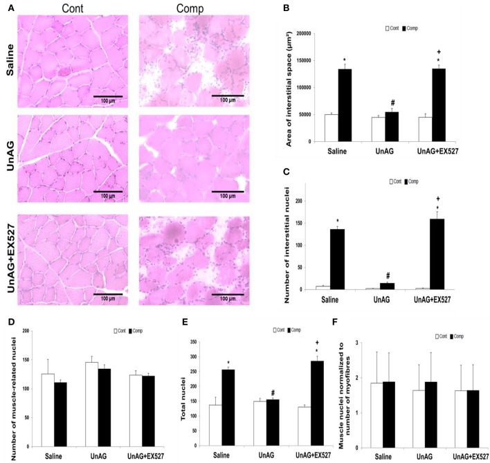
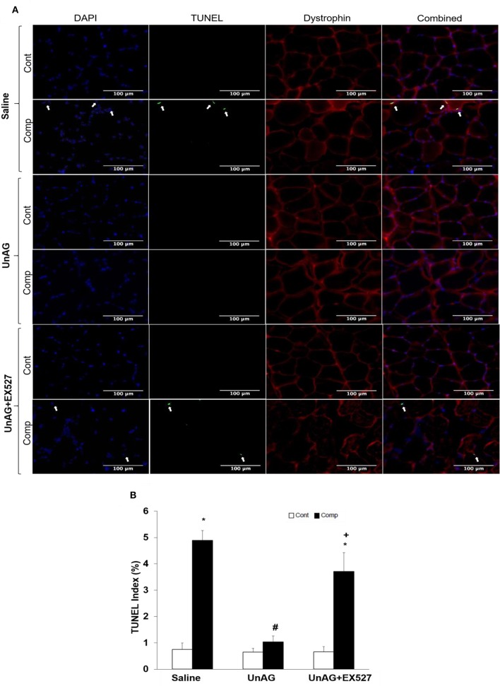
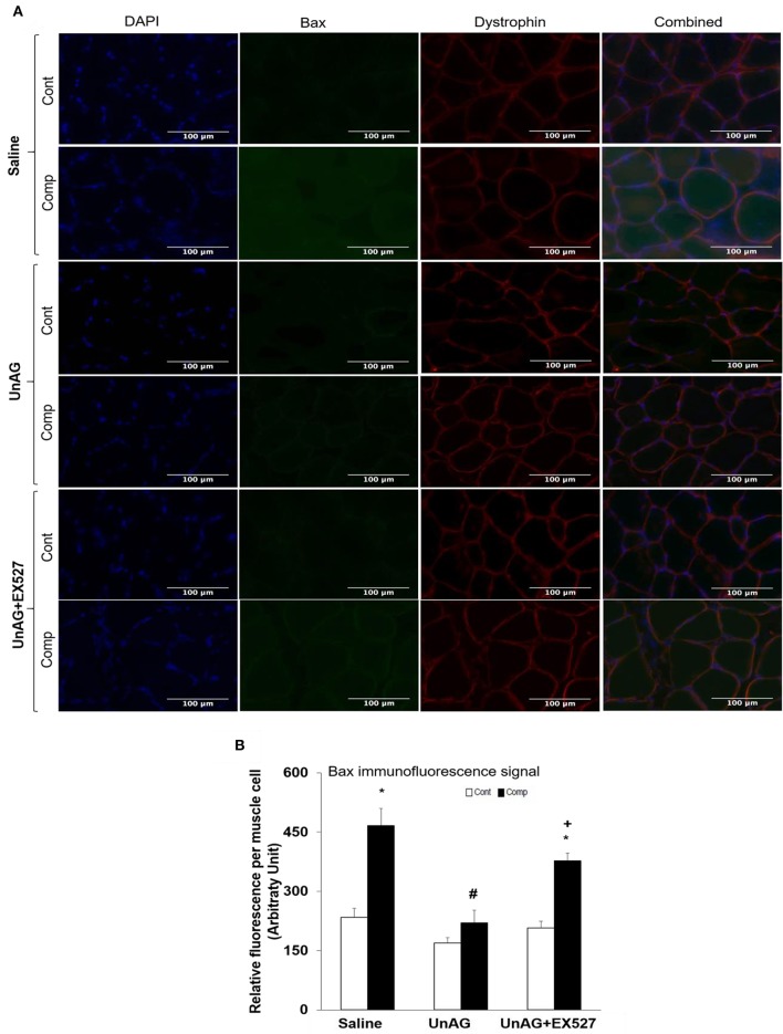
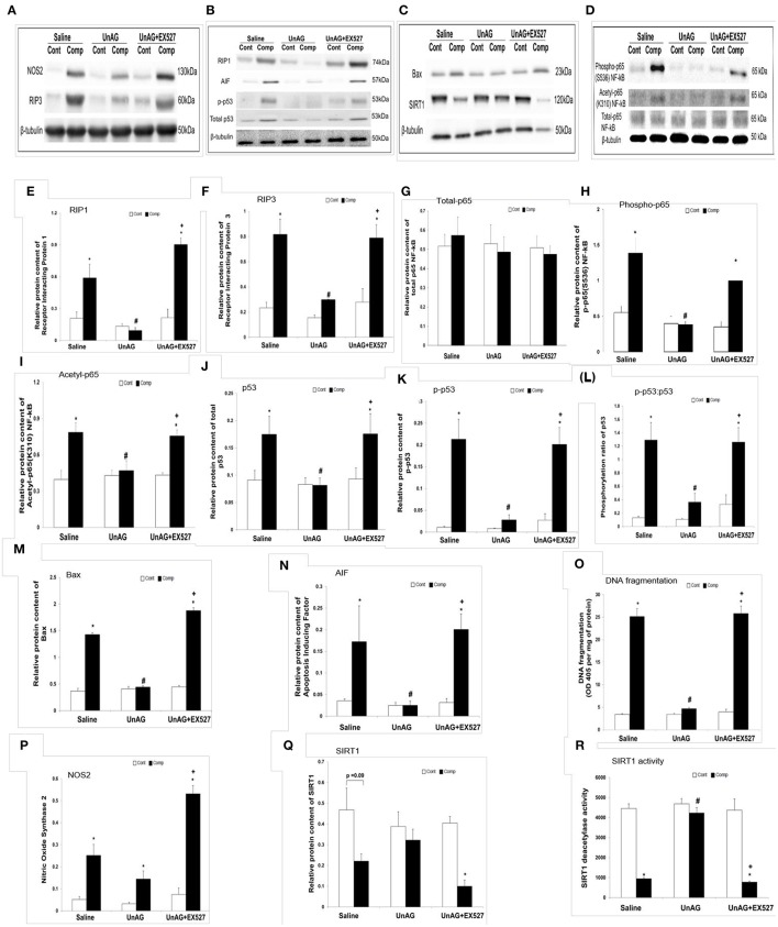
Unacylated ghrelin, the predominant form of circulating ghrelin, protects myotubes from cell death, which is a known attribute of pressure ulcers. In this study, we investigated whether unacylated ghrelin protects skeletal muscle from pressure-induced deep tissue injury by abolishing necroptosis and apoptosis signaling and whether these effects were mediated by SIRT1 pathway. Fifteen adult Sprague Dawley rats were assigned to receive saline or unacylated ghrelin with or without EX527 (a SIRT1 inhibitor). Animals underwent two 6-h compression cycles with 100 mmHg static pressure applied over the mid-tibialis region of the right limb whereas the left uncompressed limb served as the intra-animal control. Muscle tissues underneath the compression region, and at the similar region of the opposite uncompressed limb, were collected for analysis. Unacylated ghrelin attenuated the compression-induced muscle pathohistological alterations including rounding contour of myofibers, extensive nucleus accumulation in the interstitial space, and increased interstitial space. Unacylated ghrelin abolished the increase in necroptosis proteins including RIP1 and RIP3 and attenuated the elevation of apoptotic proteins including p53, Bax, and AIF in the compressed muscle. Furthermore, unacylated ghrelin opposed the compression-induced phosphorylation and acetylation of p65 subunit of NF-kB. The anti-apoptotic effect of unacylated ghrelin was shown by a decrease in apoptotic DNA fragmentation and terminal dUTP nick-end labeling index in the compressed muscle. The protective effects of unacylated ghrelin vanished when co-treated with EX527. Our findings demonstrated that unacylated ghrelin protected skeletal muscle from compression-induced injury. The myoprotective effects of unacylated ghrelin on pressure-induced tissue injury were associated with SIRT1 signaling.

Keywords: EX527; apoptosis; necroptosis; oxidative stress; pressure sores; unacylated ghrelin.


Figures

Figure 1
Figure 1
Histological analyses. UnAG abolished abnormal muscle histology induced by moderate compression but this effect was vanished when co-treated with EX527 (A). The elevation of area of interstitial space induced by compression injury was alleviated by UnAG (B). The reduction of the number of interstitial nuclei induced by UnAG in the compressed muscle was mitigated by the co-treatment of EX527 (C). No compression or treatment effect was observed for the number of muscle-related nuclei (D). The total number of nuclei was significantly different between compressed and uncompressed muscles in all groups except in the group treated only with UnAG (E). Neither compression nor drug treatment affected the number of muscle nuclei normalized to the number of myofibers (F). *p < 0.05, compressed muscle compared to uncompressed control muscle; #p < 0.05, UnAG compared to Saline; +p < 0.05, UnAG+EX527 compared to UnAG; Cont, control muscle; Comp, compressed muscle.
Figure 2
Figure 2
TUNEL and dystrophin staining. Apoptotic nuclear DNA breaks in muscle tissue were measured using the technique of TUNEL staining (A) and were expressed as TUNEL index (B). Representative images of TUNEL and dystrophin staining of compressed and uncompressed muscles (A). Immunofluorescence labeling of dystrophin (red) was performed to identify the localization of the TUNEL-positive nuclei (green) with regard to muscle sarcolemmal membrane and nuclei were labeled with DAPI (blue) (A). UnAG opposed the increase of TUNEL positive nuclei induced by moderate compression but this protective effect was absent when co-treated with EX527 (A). TUNEL index was higher in compressed muscles of saline and UnAG+EX527 groups compared to compressed muscle of UnAG group (B). Arrows indicate TUNEL-positive nuclei. *p < 0.05, compressed muscle compared to uncompressed control muscle; #p < 0.05, UnAG compared to Saline; +p < 0.05, UnAG+EX527 compared to UnAG; Cont, control muscle; Comp, compressed muscle; DAPI, 4′,6-diamidino-2-phenyl-lindole.
Figure 3
Figure 3
Immunofluorescence staining of Bax and dystrophin. Representative images of compressed and uncompressed muscles labeled with anti-dystrophin (red) and anti-bax (green) (A). Muscle Bax signal was increased in compressed muscles treated with saline and UnAG+EX527 (A). In contrast, muscle Bax abundance was decreased in compressed muscles treated with only UnAG (A). Quantification of relative fluorescence intensity of Bax (B). *p < 0.05, compressed muscle compared to uncompressed control muscle; #p < 0.05, UnAG compared to Saline; +p < 0.05, UnAG+EX527 compared to UnAG; Cont, control muscle; Comp, compressed muscle.
Figure 4
Figure 4
Immunoblot and biochemical analyses. For the immunoblotting, all samples were probed for the examined protein with the respective antibody on the same membrane which underwent the same probing and washing procedure. The representative blot pictures are shown in (A–D). UnAG, but not in conjunction with EX527, blunted the increase in protein abundance of RIP1 in compressed muscle (E). Similar pattern was observed in the protein abundance of RIP3 (F). Total p65 NF-kB protein abundance did not change in the compressed muscle when compared to uncompressed muscle in all groups (G). The immunoblot analyses revealed the suppressive effects of UnAG, but not in conjunction with EX527, on the abundances of phospho-p65 NF-kB (H), acetyl-p65 NF-kB (I), p53 (J), phospho-p53 (K), phosphorylation ratio of p53 (L), Bax (M), and AIF (N). The anti-apoptotic effect of UnAG was confirmed by apoptotic DNA fragmentation index (O). The protein content of NOS2 was significantly elevated in the compressed muscles in all groups, irrespective of drug administered (P). SIRT1 protein abundance was tended to decrease in the compressed muscle when compared to uncompressed muscle in saline group (Q). SIRT1 protein abundance was significantly decreased in muscle co-treated with UnAG and EX527 after compression (Q). The deacetylase activity of SIRT1 was significantly decreased after compression in all groups except in compressed muscle treated only with UnAG (R). *p < 0.05, compressed muscle compared to uncompressed control muscle; #p < 0.05, UnAG compared to Saline; +p < 0.05, UnAG+EX527 compared to UnAG; Cont, control muscle; Comp, compressed muscle.

References

    1. Agrawal K., Chauhan N. (2012). Pressure ulcers: back to the basics. Indian J. Plast. Surg. 45, 244–254. 10.4103/0970-0358.101287 - DOI - PMC - PubMed
    1. Bagul P. K., Deepthi N., Sultana R., Banerjee S. K. (2015). Resveratrol ameliorates cardiac oxidative stress in diabetes through deacetylation of NFkB-p65 and histone 3. J. Nutr. Biochem. 26, 1298–1307. 10.1016/j.jnutbio.2015.06.006 - DOI - PubMed
    1. Bauer K., Rock K., Nazzal M., Jones O., Qu W. (2016). Pressure ulcers in the United States' inpatient population from 2008 to 2012: results of a retrospective nationwide study. Ostomy Wound Manage. 62, 30–38. - PubMed
    1. Beckman J. S., Beckman T. W., Chen J., Marshall P. A., Freeman B. A. (1990). Apparent hydroxyl radical production by peroxynitrite: implications for endothelial injury from nitric oxide and superoxide. Proc. Natl. Acad. Sci. U.S.A. 87, 1620–1624. 10.1073/pnas.87.4.1620 - DOI - PMC - PubMed
    1. Bennett G., Dealey C., Posnett J. (2004). The cost of pressure ulcers in the UK. Age Ageing 33, 230–235. 10.1093/ageing/afh086 - DOI - PubMed