. 2016 Apr;22(4):142-151.
doi: 10.1016/j.mpdhp.2016.04.004. Epub 2016 May 21.

Unusual variants of mycosis fungoides

Affiliations

Unusual variants of mycosis fungoides

Pooja Virmani et al. Diagn Histopathol (Oxf). 2016 Apr.

Abstract

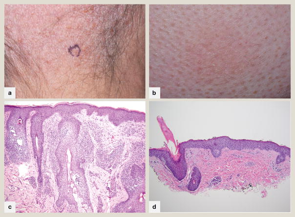
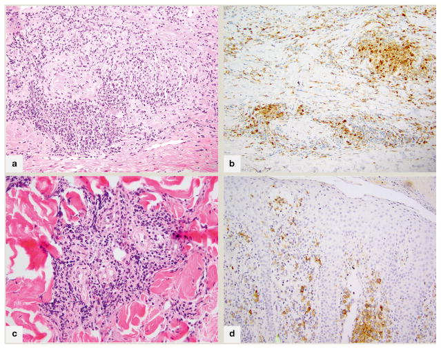
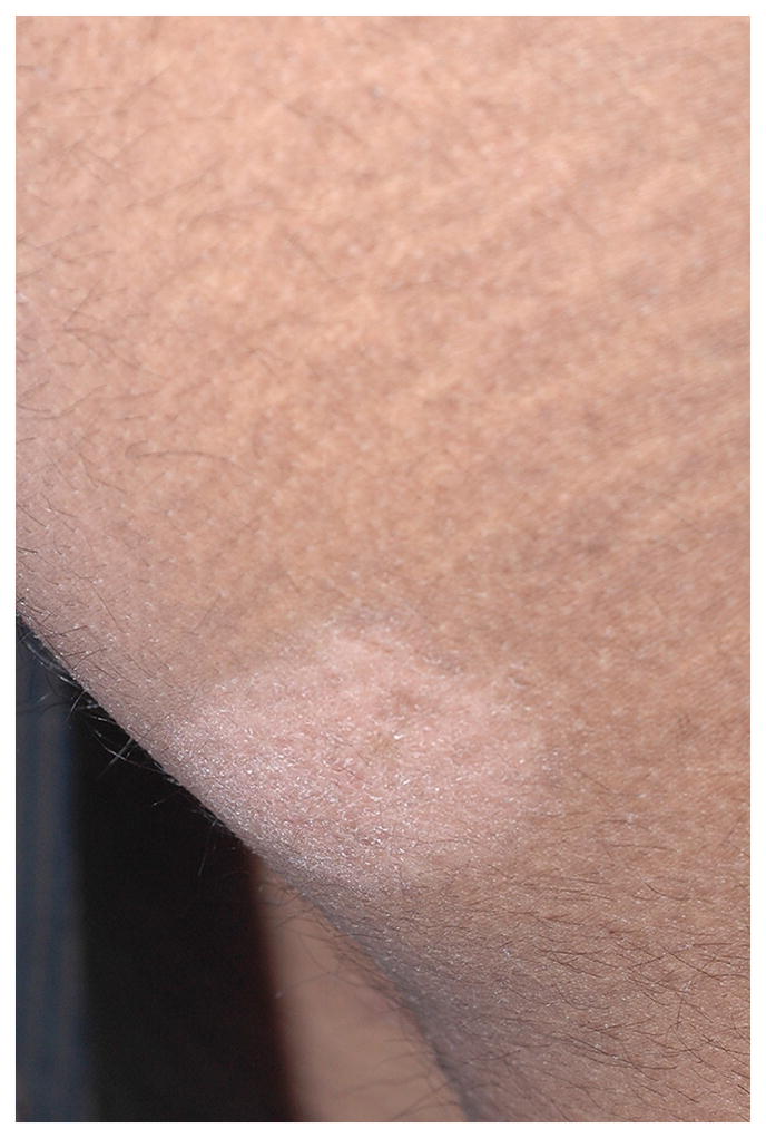
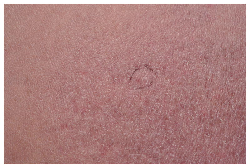
Conventional presentations of mycosis fungoides may be diagnostically challenging, particularly in light of the controversial boundaries defining the disease. Variant presentations of this cutaneous T-cell lymphoma add a further layer of complexity, requiring a sophisticated and informed perspective when evaluating lymphoid infiltrates in the skin. Herein we discuss well-defined (WHO-EORTC) variants pagetoid reticulosis, granulomatous slack skin and folliculotropic mycosis fungoides as well as less well-defined morphologic/architectural variants, and divergent immunohistochemical presentations of this typically indolent T-cell lymphoproliferative disease.

Keywords: Cutaneous T-cell lymphoma; granulomatous slack skin; lymphoproliferative disorders; mycosis fungoides; non-Hodgkin; pagetoid reticulosis; peripheral T-cell lymphoma.


Conflict of interest statement

Conflicts of interest: none declared.

Figures

Fig 1
Fig 1
Folliculotropic mycosis fungoides A. Alopetic patch on scalp revealing patterned follicle-centered erythema with collarettes of scale. B. Perifollicular erythema and scale with markedly hyperkeratotic follicular plugs in a patient with spiky folliculotropic mycosis fungoides. C. 100x magnification. Atypical lymphocytes extend exuberantly into follicular epithelium, sparing the overlying epidermis with follicular dyskeratosis typical of folliculotropic mycosis fungoides D. 100x magnification. In this photomicrograph of spiky mycosis fungoides, atypical lymphocytes are present within follicular epithelium below a hyperkeratotic column emanating from the hair follicle.
Fig 2
Fig 2
Fig 2A. 200x magnification; granulomatous mycosis fungoides with prominent granuloma annulare-like appearance (palisading of histiocytes within papillary dermis), associated with atypical lymphocytes B. 200x magnification; an immunohistochemical stain for CD68 highlights the 25% component of histiocytes in this case C. 200x magnification; in syringotropic mycosis fungoides, a dense lymphoid infiltrate surrounds the eccrine glands and extends into glandular epithelium D. CD30 immunohistochemical stain, 200x; CD30+ mycosis fungoides with two distinct populations of lymphocytes characterized by CD30 positivity and small (intraepidermal) or large(dermal) cell size.
Fig 3
Fig 3
Hypopigmented mycosis fungoides; a typical hypopigmented to achromic patch on the buttock
Fig 4
Fig 4
Poikilodermatous mycosis fungoides; an atrophic patch showing both hypo- and hyperpigmentation with telangiectasia
Fig 5
Fig 5
CD8+ mycosis fungoides A. 400x magnification of an acanthotic lesion with prominent basal layer tagging and marked extension of lymphocytes into mid to upper layers of epidermis. A few necrotic keratinocytes are present B. 400x magnification of an atrophic lesion with marked basilar epidermotropism. Both A and B show few remaining pigmented basilar keratinocytes, but many superficial dermal melanophages, connoting basal layer destruction and melanin dropout. C. 200x CD8 immunohistochemical stain of case in part A showing diffuse staining of all epidermotropic lymphocytes D. 200x CD7 immunohistochemical stain of case in part A shows characteristic complete absence of this pan T-cell antigen

References

    1. Willemze R, et al. WHO-EORTC classification for cutaneous lymphomas. Blood. 2005;105:3768–3785. - PubMed
    1. Agar NS, et al. Survival outcomes and prognostic factors in mycosis fungoides/Sezary syndrome: validation of the revised International Society for Cutaneous Lymphomas/European Organisation for Research and Treatment of Cancer staging proposal. J Clin Oncol. 2010;28:4730–4739. - PubMed
    1. Olsen E, et al. Revisions to the staging and classification of mycosis fungoides and Sezary syndrome: a proposal of the International Society for Cutaneous Lymphomas (ISCL) and the cutaneous lymphoma task force of the European Organization of Research and Treatment of Cancer (EORTC) Blood. 2007;110:1713–1722. - PubMed
    1. Haghighi B, et al. Pagetoid reticulosis (Woringer-Kolopp disease): an immunophenotypic, molecular, and clinicopathologic study. Mod Pathol. 2000;13:502–510. - PubMed
    1. Rodriguez-Pinilla SM, et al. TCR-gamma expression in primary cutaneous T-cell lymphomas. Am J Surg Pathol. 2013;37:375–384. - PubMed

LinkOut - more resources