Review
. 2017:2017:5293258.
doi: 10.1155/2017/5293258. Epub 2017 Nov 1.

Cellular Senescence in Age-Related Macular Degeneration: Can Autophagy and DNA Damage Response Play a Role?

Affiliations
Review

Cellular Senescence in Age-Related Macular Degeneration: Can Autophagy and DNA Damage Response Play a Role?

Janusz Blasiak et al. Oxid Med Cell Longev. 2017.

Abstract

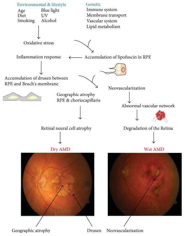
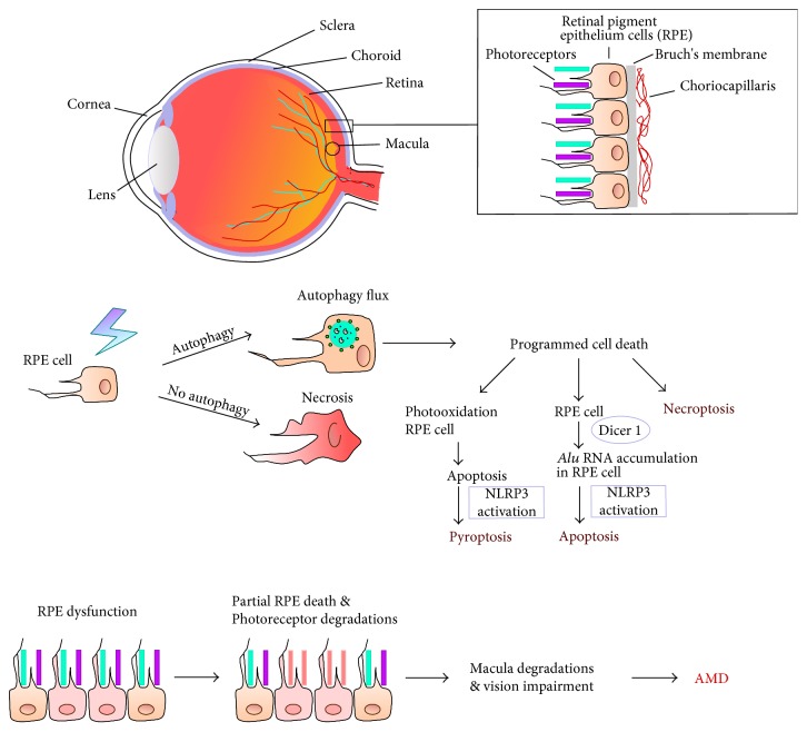
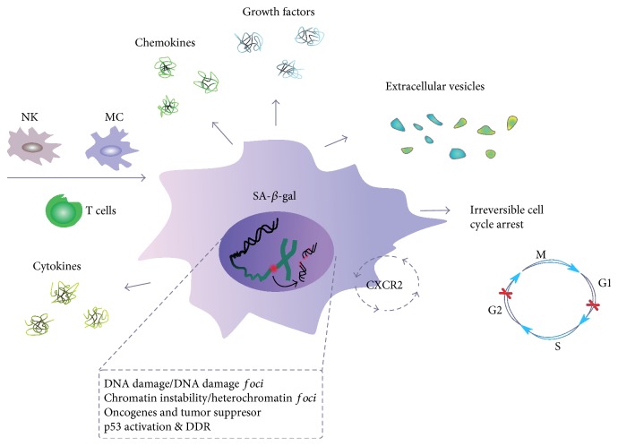
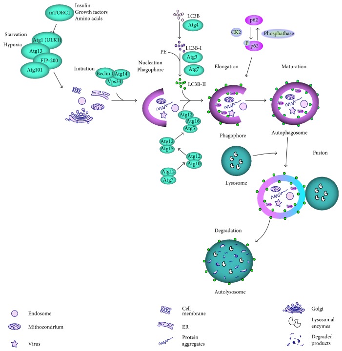
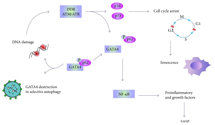
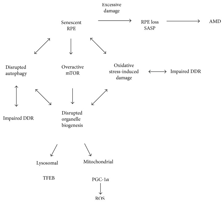
Age-related macular degeneration (AMD) is the main reason of blindness in developed countries. Aging is the main AMD risk factor. Oxidative stress, inflammation and some genetic factors play a role in AMD pathogenesis. AMD is associated with the degradation of retinal pigment epithelium (RPE) cells, photoreceptors, and choriocapillaris. Lost RPE cells in the central retina can be replaced by their peripheral counterparts. However, if they are senescent, degenerated regions in the macula cannot be regenerated. Oxidative stress, a main factor of AMD pathogenesis, can induce DNA damage response (DDR), autophagy, and cell senescence. Moreover, cell senescence is involved in the pathogenesis of many age-related diseases. Cell senescence is the state of permanent cellular division arrest and concerns only mitotic cells. RPE cells, although quiescent in the retina, can proliferate in vitro. They can also undergo oxidative stress-induced senescence. Therefore, cellular senescence can be considered as an important molecular pathway of AMD pathology, resulting in an inability of the macula to regenerate after degeneration of RPE cells caused by a factor inducing DDR and autophagy. It is too early to speculate about the role of the mutual interplay between cell senescence, autophagy, and DDR, but this subject is worth further studies.


Figures

Figure 1
Figure 1
The exact mechanism of AMD pathogenesis is not known, but several factors can be implicated with a distinct role of aging. Besides aging, various oxidative stress-related environmental and lifestyle influences can be involved. The complement gene mutations play a major role in AMD. Oxidative stress and presumably other factors lead to accumulation of heterogenous lysosomal lipofuscin in retinal pigment epithelium (RPE), which induces a proinflammatory response. This, in turn, can lead to accumulation of extracellular drusen. Lipofuscin contains proangiogenic factors, such as A2E, that may develop choroidal neovascularization typical for wet AMD.
Figure 2
Figure 2
Cell death and autophagy in AMD progression. AMD affects the macula, a part in the central retina, and is associated with degradation of retinal pigment epithelium (RPE) cells, photoreceptors, and choriocapillaris. Autophagy can be decisive in switching between programmed and nonprogrammed cell death mode. Apoptosis of RPE cells can be linked to blue light exposure (photooxidation), oxidative stress, accumulation of Alu transposons due to impaired functioning of the DICER1 endonuclease, and the activation of the NLRP3 inflammasome. Pyroptosis can also result from photooxidation and activation of the NLRP3 inflammasome. Oxidative stress and other factors can induce necroptosis, a programmed version of necrosis.
Figure 3
Figure 3
Senescent cells and senescence-associated secretory phenotype (SASP). A cell undergoing senescence is permanently arrested in the G1 or G2 phase of the cell cycle and has changed morphology. It is featured by an increased activity of senescence-associated-β-galactosidase (SA-β-gal) and can be targeted by the immunological system, with natural killer (NK) cells, macrophages (MS), and T-lymphocytes involved. Released various soluble agents, including cytokines, chemokines, growth factors, and extracellular vesicles, are main determinants of SASP. A senescent cell is characterized by an elevated level of DNA damage and chromosomes aberrations, which are also signs of genomic and chromosomal instability, typical for cancer cells. Chemokine signaling through the CXCR2 protein increases senescence.
Figure 4
Figure 4
Autophagy dependent on mTOR. In normal nutrient conditions, the mTOR complex 1 (mTORC1) inhibits the ULK1 complex, consisting of ULK1, Atg13, Atg101, and FIP200, which can activate autophagy in stress conditions, including starvation and hypoxia or when the inhibitory effect of mTORC1 is abolished by growth factors, insulin, amino acids, or other agents. The material to be degraded (cargo) is then enclosed by a nucleating phagophore, which requires a translocation of ULK1 to endoplasmic reticulum (ER). ER membrane is used to form the phagophore, but other sources are also possible. The phagophore membrane is elongated, which leads to the formation of autophagosome, a vesicle with the enclosed cargo. This process is assisted by LC3 lipidated by phosphatidylethanolamine (PE) and many individual proteins, including Beclin 1, Vps34, and autophagy-related proteins (Atgs). The p62 protein functions as a selective autophagy receptor for degradation of ubiqutinated substrates, but it is itself a specific substrate for autophagy after its phosphorylation and can be selectively incorporated into the autophagosome and degraded. Fusion of autophagosome with lysosome creates autolysosome in which the cargo is degraded by lysosomal enzymes. Autophagy can be also activated by mTOR-independent pathways.
Figure 5
Figure 5
Nuclear and mitochondrial DNA (nDNA and mtDNA) can be damaged by AMD risk factors, which can also affect proteins, including DNA repair proteins. Nonrepaired or misrepaired DNA can contribute to retinal cell death occurring in AMD [89].
Figure 6
Figure 6
GATA4 can be involved in autophagy, senescence, and DNA damage response (DDR). The level of GATA4 is normally regulated by p62-dependent selective autophagy, but DNA damage and resulting DDR can release GATA4 from p62 control by its ATM-induced phosphorylation. If DNA damage cannot be repaired, DDR effectors induce permanent and irreversible cell cycle arrest, which is a hallmark of senescence with senescence-associated phenotype (SASP). GATA4 released from autophagic degradation can transactivate several genes that activate NF-κB, resulting in the release of growth factors, chemokines, cytokines, and other molecules typical for SASP.
Figure 7
Figure 7
Senescence as a critical factor in AMD pathogenesis. In certain stress conditions, which can be induced by environmental or/and lifestyle factors in aging retina, a major fraction of RPE cells become senescent and are no longer able to regenerate damaged RPE cells, which leads to AMD. The senescence of RPE cells can result from an interplay between aging, autophagy, and DDR in stress conditions. This interplay is a kind of vicious cycle as impaired DNA damage response (DDR) can lead to an increased damage to biomolecules by ROS. Damage to biomolecules induces the degradation of organelles via mTOR-dependent autophagy. This may lead to aggravation of oxidative stress and cellular damage as well as continue to impair autophagy and antioxidant defense by altered TFEB (transcription factor E-box binding) and PGC-1α signaling and increased ROS generation.

References

    1. Wong W. L., Su X., Li X., et al. Global prevalence of age-related macular degeneration and disease burden projection for 2020 and 2040: a systematic review and meta-analysis. The Lancet Global Health. 2014;2(2):e106–e116. doi: 10.1016/S2214-109X(13)70145-1. - DOI - PubMed
    1. Jonas J. B. Global prevalence of age-related macular degeneration. The Lancet Global Health. 2014;2(2):e65–e66. doi: 10.1016/S2214-109X(13)70163-3. - DOI - PubMed
    1. Gordois A., Cutler H., Pezzullo L., et al. An estimation of the worldwide economic and health burden of visual impairment. Global Public Health. 2012;7(5):465–481. doi: 10.1080/17441692.2011.634815. - DOI - PubMed
    1. Reibaldi M., Longo A., Pulvirenti A., et al. Geo-epidemiology of age-related macular degeneration: new clues into the pathogenesis. American Journal of Ophthalmology. 2016;161:78.e1–93.e2. doi: 10.1016/j.ajo.2015.09.031. - DOI - PubMed
    1. Kauppinen A., Paterno J. J., Blasiak J., Salminen A., Kaarniranta K. Inflammation and its role in age-related macular degeneration. Cellular and Molecular Life Sciences. 2016;73(9):1765–1786. doi: 10.1007/s00018-016-2147-8. - DOI - PMC - PubMed

MeSH terms

Substances