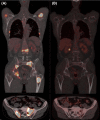
Multiple large osteolytic lesions in a patient with systemic mastocytosis: a challenging diagnosis
- PMID: 29225841
- PMCID: PMC5715414
- DOI: 10.1002/ccr3.1232
Multiple large osteolytic lesions in a patient with systemic mastocytosis: a challenging diagnosis
Abstract
Patients with advanced variants of Systemic Mastocytosis may develop destructive bone lesions when massive mast cell (MC) infiltrates are present. Finding of large osteolyses in indolent systemic mastocytosis, typically characterized by low MC burden, should prompt investigations for an alternative explanation.
Keywords: Non‐hodgkin lymphoma; osteolysis; primary bone lymphoma; systemic mastocytosis; tryptase.
Figures


References
-
- Arber, D. A. , Orazi A., Hasserjian R., Thiele J., Borowitz M. J., and Le Beau M. M., et al. 2016. The 2016 revision to the World Health Organization classification of myeloid neoplasms and acute leukemia. Blood 127:2391–2405. - PubMed
-
- Lim, K. H. , Tefferi A., Lasho T. L., Finke C., Patnaik M., and Butterfield J. H., et al. 2009. Systemic mastocytosis in 342 consecutive adults: survival studies and prognostic factors. Blood 113:5727–5736. - PubMed
-
- Barete, S. , Assous N., de Gennes C., Grandpeix C., Feger F., and Palmerini F., et al. 2010. Systemic mastocytosis and bone involvement in a cohort of 75 patients. Ann. Rheum. Dis. 69:1838–1841. - PubMed
-
- Rossini, M. , Zanotti R., Orsolini G., Tripi G., Viapiana O., and Idolazzi L., et al. 2016. Prevalence, pathogenesis, and treatment options for mastocytosis‐related osteoporosis. Osteoporos. Int. 27:2411–2421. - PubMed
-
- Escribano, L. , Alvarez‐Twose I., Sánchez‐Muñoz L., Garcia‐Montero A., Nunez R., and Almeida J., et al. 2009. Prognosis in adult indolent systemic mastocytosis: a long‐term study of the Spanish Network on Mastocytosis in a series of 145 patients. J. Allergy Clin. Immunol. 124:514–521. - PubMed
Publication types
LinkOut - more resources
Full Text Sources
Other Literature Sources

